Revision 1
#96396
Store at -20C
877-616-CELL (2355)
877-678-TECH (8324)
3 Trask Lane | Danvers | Massachusetts | 01923 | USA
For Research Use Only. Not for Use in Diagnostic Procedures.
Applications:
W, IP, IF-F, IF-IC
Reactivity:
H M R
Sensitivity:
Endogenous
MW (kDa):
90
Source/Isotype:
Rabbit IgG
UniProt ID:
#O94874
Entrez-Gene Id:
23376
Product Usage Information
| Application | Dilution |
|---|---|
| Western Blotting | 1:1000 |
| Immunoprecipitation | 1:50 |
| Immunofluorescence (Frozen) | 1:100 - 1:400 |
| Immunofluorescence (Immunocytochemistry) | 1:100 - 1:200 |
Storage
Specificity/Sensitivity
UFL1 (F5P6L) Rabbit mAb recognizes endogenous levels of total UFL1 protein.
Source / Purification
Monoclonal antibody is produced by immunizing animals with a synthetic peptide corresponding to residues near the carboxy terminus of human UFL1 protein.
Background
The ubiquitin-fold modifier 1 (UFM1) conjugation system (UFMylation) is a ubiquitin-like system that plays key roles in development and stress responses (1, reviewed in 2-4). Like ubiquitin, conjugation of UFM1 is a three-step enzymatic process involving a specific E1-activating enzyme (UBA5), an E2-conjugating enzyme (UFC1), and an E3-ligation enzyme (UFL1). Several substrates for UFMylation have been identified including DDRGK1, RPL26, p53, MRE11, histone H4, ASC1, CDK5RAP3, and CYB5R3 (5-12). Modification of these targets by UFM1 has direct effects on ER homeostasis, protein translation, and response to DNA damage. Notably, independent screens identified UFMylation as a key regulator of ER-phagy and autophagy (5,13,14). Aberrant UFMylation is associated with many pathological conditions, including cancer, diabetes, and inflammatory diseases (2-4).
Background References
- Tatsumi, K. et al. (2010) J Biol Chem 285, 5417-27.
- Gerakis, Y. et al. (2019) Trends Cell Biol 29, 974-986.
- Jiang, Q. et al. (2023) Front Endocrinol (Lausanne) 14, 1123124.
- Jing, Y. et al. (2022) Cancers (Basel) 14, 3501.
- Liu, J. et al. (2017) Nat Commun 8, 14186.
- Walczak, C.P. et al. (2019) Proc Natl Acad Sci USA 116, 1299-1308.
- Liu, J. et al. (2020) Nat Cell Biol 22, 1056-1063.
- Lee, L. et al. (2021) Sci Adv 7, eabc7371.
- Qin, B. et al. (2019) Nat Commun 10, 1242.
- Yoo, H.M. et al. (2014) Mol Cell 56, 261-274.
- Yang, R. et al. (2019) Development 146, dev169235. doi: 10.1242/dev.169235.
- Ishimura, R. et al. (2022) Nat Commun 13, 7857.
- Liang, J.R. et al. (2020) Cell 180, 1160-1177.e20.
- DeJesus, R. et al. (2016) Elife 5, e17290. doi: 10.7554/eLife.17290.
Species Reactivity
Species reactivity is determined by testing in at least one approved application (e.g., western blot).
Western Blot Buffer
IMPORTANT: For western blots, incubate membrane with diluted primary antibody in 5% w/v nonfat dry milk, 1X TBS, 0.1% Tween® 20 at 4°C with gentle shaking, overnight.
Applications Key
W: Western Blotting IP: Immunoprecipitation IF-F: Immunofluorescence (Frozen)
Cross-Reactivity Key
H: Human M: Mouse R: Rat Hm: Hamster Mk: Monkey Vir: Virus Mi: Mink C: Chicken Dm: D. melanogaster X: Xenopus Z: Zebrafish B: Bovine Dg: Dog Pg: Pig Sc: S. cerevisiae Ce: C. elegans Hr: Horse GP: Guinea Pig Rab: Rabbit G: Goat All: All Species Expected
Trademarks and Patents
Cell Signaling Technology is a trademark of Cell Signaling Technology, Inc.
All other trademarks are the property of their respective owners. Visit cellsignal.com/trademarks for more information.
Limited Uses
Except as otherwise expressly agreed in a writing signed by a legally authorized representative of CST, the following terms apply to Products provided by CST, its affiliates or its distributors. Any Customer's terms and conditions that are in addition to, or different from, those contained herein, unless separately accepted in writing by a legally authorized representative of CST, are rejected and are of no force or effect.
Products are labeled with For Research Use Only or a similar labeling statement and have not been approved, cleared, or licensed by the FDA or other regulatory foreign or domestic entity, for any purpose. Customer shall not use any Product for any diagnostic or therapeutic purpose, or otherwise in any manner that conflicts with its labeling statement. Products sold or licensed by CST are provided for Customer as the end-user and solely for research and development uses. Any use of Product for diagnostic, prophylactic or therapeutic purposes, or any purchase of Product for resale (alone or as a component) or other commercial purpose, requires a separate license from CST. Customer shall (a) not sell, license, loan, donate or otherwise transfer or make available any Product to any third party, whether alone or in combination with other materials, or use the Products to manufacture any commercial products, (b) not copy, modify, reverse engineer, decompile, disassemble or otherwise attempt to discover the underlying structure or technology of the Products, or use the Products for the purpose of developing any products or services that would compete with CST products or services, (c) not alter or remove from the Products any trademarks, trade names, logos, patent or copyright notices or markings, (d) use the Products solely in accordance with CST Product Terms of Sale and any applicable documentation, and (e) comply with any license, terms of service or similar agreement with respect to any third party products or services used by Customer in connection with the Products.
Revision 1
#96396
UFL1 (F5P6L) Rabbit mAb
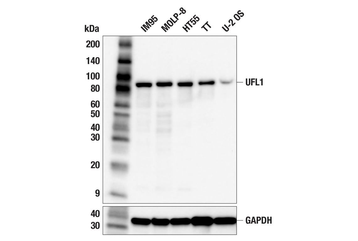
Western blot analysis of extracts from various cell lines using UFL1 (F5P6L) Rabbit mAb (upper) or GAPDH (D16H11) XP® Rabbit mAb #5174 (lower). Low expression of UFL1 protein in U-2 OS cells is consistent with the predicted expression pattern.
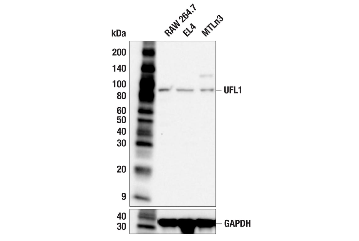
Western blot analysis of extracts from RAW 264.7, EL4, and MTLn3 cells using UFL1 (F5P6L) Rabbit mAb (upper) or GAPDH (D16H11) XP® Rabbit mAb #5174 (lower).
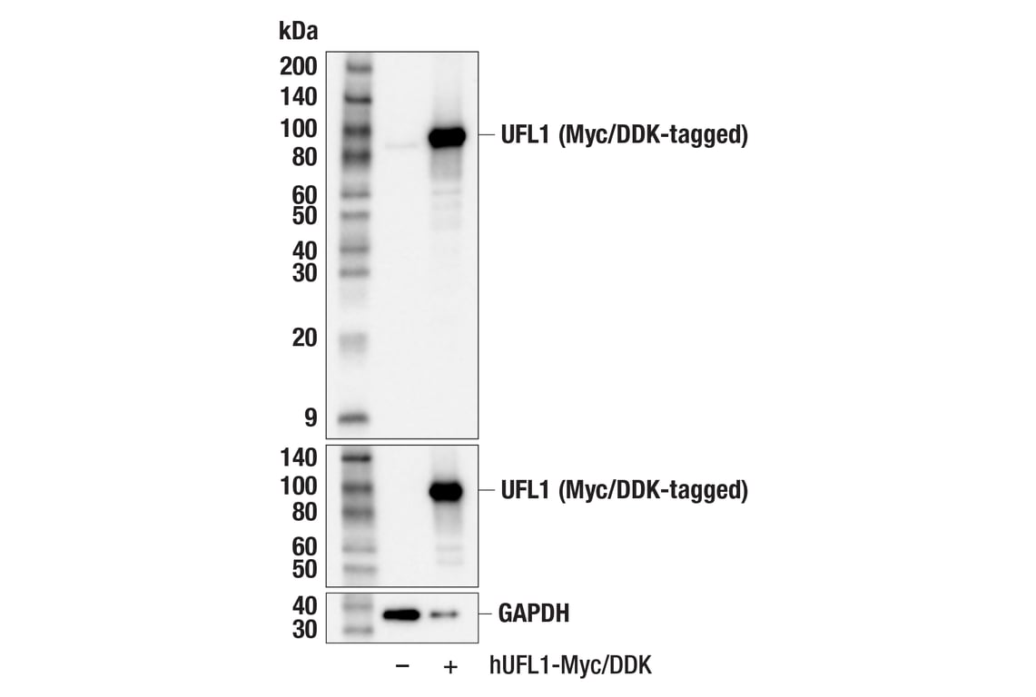
Western blot analysis of extracts from 293T cells, mock transfected (-) or transfected with a construct expressing Myc/DDK-tagged full-length human UFL1 protein (hUFL1-Myc/DDK; +), using UFL1 (F5P6L) Rabbit mAb (upper), Myc-Tag (71D10) Rabbit mAb #2278 (middle), or GAPDH (D16H11) XP® Rabbit mAb #5174 (lower).
Revision 1
#96396
UFL1 (F5P6L) Rabbit mAb
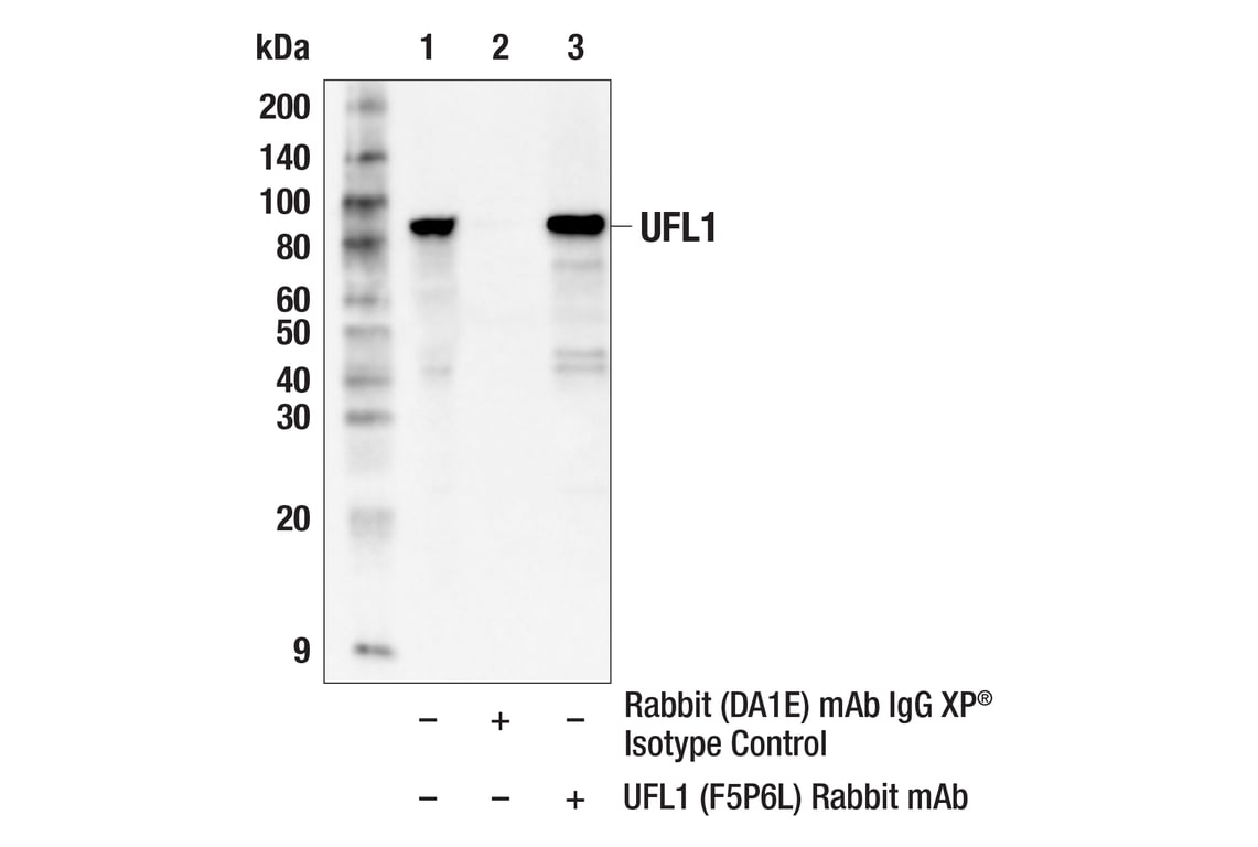
Immunoprecipitation of UFL1 protein from HT55 cell extracts. Lane 1 is 10% input, lane 2 is Rabbit (DA1E) mAb IgG XP® Isotype Control #3900, and lane 3 is UFL1 (F5P6L) Rabbit mAb. Western blot analysis was performed using UFL1 (F5P6L) Rabbit mAb. Mouse Anti-rabbit IgG (Conformation Specific) (L27A9) mAb (HRP Conjugate) #5127 was used as a secondary antibody.
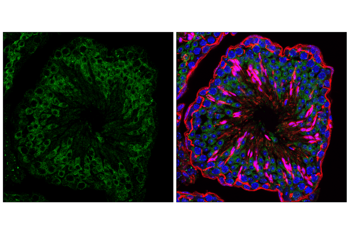
Confocal immunofluorescent analysis of fixed frozen mouse testis labeled with UFL1 (F5P6L) Rabbit mAb (green), DyLight 650 Phalloidin #12956 (red), and ProLong Gold Antifade Reagent with DAPI #8961 (blue).
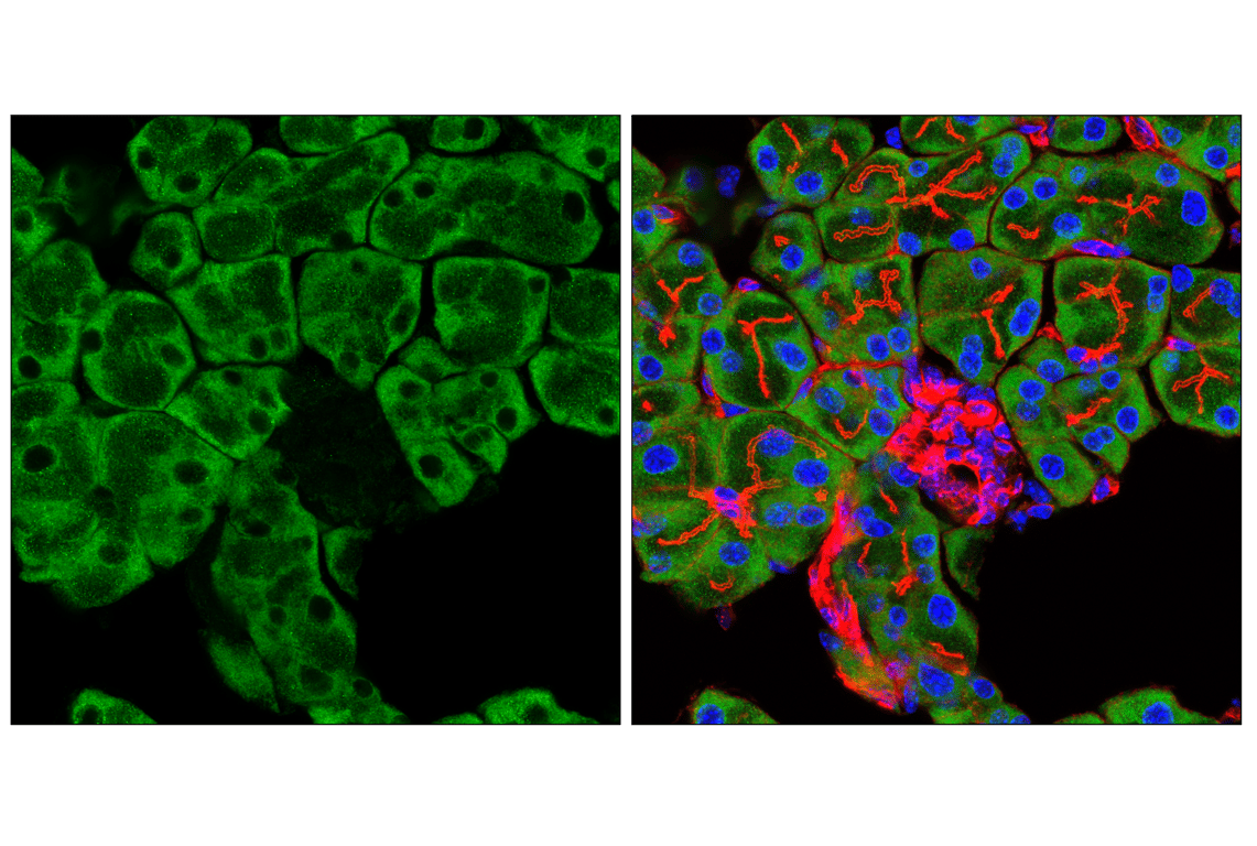
Confocal immunofluorescent analysis of fixed frozen mouse pancreas, labeled with UFL1 (F5P6L) Rabbit mAb (left, green) and co-labeled with DyLight 650 Phalloidin #12956 (right, red) and ProLong Gold Antifade Reagent with DAPI #8961 (right, blue).
Revision 1
#96396
UFL1 (F5P6L) Rabbit mAb
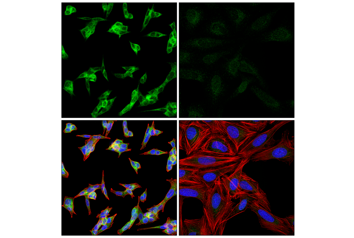
Confocal immunofluorescent analysis of TT cells (left, high-expressing) and U-2 OS cells (right, low-expressing) using UFL1 (F5P6L) Rabbit mAb (green), DyLight 554 Phalloidin #13054 (red), and DAPI #4083 (blue).