Revision 1
#94886
Store at -20C
877-616-CELL (2355)
877-678-TECH (8324)
3 Trask Lane | Danvers | Massachusetts | 01923 | USA
For Research Use Only. Not for Use in Diagnostic Procedures.
Applications:
W, IP, IF-IC
Reactivity:
H
Sensitivity:
Endogenous
MW (kDa):
79
Source/Isotype:
Rabbit IgG
UniProt ID:
#O15297
Entrez-Gene Id:
8493
Product Usage Information
| Application | Dilution |
|---|---|
| Western Blotting | 1:1000 |
| Immunoprecipitation | 1:100 |
| Immunofluorescence (Immunocytochemistry) | 1:800 - 1:3200 |
Storage
Specificity/Sensitivity
WIP1 (E2X1I) Rabbit mAb recognizes endogenous levels of total WIP1 protein.
Source / Purification
Monoclonal antibody is produced by immunizing animals with a synthetic peptide corresponding to residues surrounding Gly383 of human WIP1 protein.
Background
Wild-type p53 induced phosphatase 1 (WIP1)/protein phosphatase magnesium-dependent 1 delta (ppm1d) is a member of the PP2C family of serine/threonine protein phosphatases. WIP1 was initially identified as a p53 target gene, induced in response to ionizing radiation (1). Studies have shown that WIP1 is overexpressed in human cancers and is involved in the regulation of multiple DNA damage signaling pathways (reviewed in 2,3). WIP1 functions in returning cells to a homeostatic state following DNA damage (4,5), as well as in maintaining p53-dependent homeostasis under non-stressed conditions (6). Researchers have shown that increased expression of WIP1 is associated with poor prognosis and lower survival rate in some human cancers (7,8). In contrast, overexpression of WIP1 in p53-negative tumor cells sensitizes them to chemotherapy-induced apoptosis while protecting normal tissue during treatment (9).
Background References
- Fiscella, M. et al. (1997) Proc Natl Acad Sci U S A 94, 6048-53.
- Zhu, Y.H. and Bulavin, D.V. (2012) Prog Mol Biol Transl Sci 106, 307-25.
- Lu, X. et al. (2008) Cancer Metastasis Rev 27, 123-35.
- Lu, X. et al. (2005) Genes Dev 19, 1162-74.
- Cha, H. et al. (2010) Cancer Res 70, 4112-22.
- Park, H.K. et al. (2011) Cell Cycle 10, 2574-82.
- Liang, C. et al. (2012) Brain Res 1444, 65-75.
- Satoh, N. et al. (2011) Cancer Sci 102, 1101-6.
- Goloudina, A.R. et al. (2012) Proc Natl Acad Sci U S A 109, E68-75.
Species Reactivity
Species reactivity is determined by testing in at least one approved application (e.g., western blot).
Western Blot Buffer
IMPORTANT: For western blots, incubate membrane with diluted primary antibody in 5% w/v BSA, 1X TBS, 0.1% Tween® 20 at 4°C with gentle shaking, overnight.
Applications Key
W: Western Blotting IP: Immunoprecipitation IF-IC: Immunofluorescence (Immunocytochemistry)
Cross-Reactivity Key
H: Human M: Mouse R: Rat Hm: Hamster Mk: Monkey Vir: Virus Mi: Mink C: Chicken Dm: D. melanogaster X: Xenopus Z: Zebrafish B: Bovine Dg: Dog Pg: Pig Sc: S. cerevisiae Ce: C. elegans Hr: Horse GP: Guinea Pig Rab: Rabbit G: Goat All: All Species Expected
Trademarks and Patents
Cell Signaling Technology is a trademark of Cell Signaling Technology, Inc.
XP is a registered trademark of Cell Signaling Technology, Inc.
All other trademarks are the property of their respective owners. Visit cellsignal.com/trademarks for more information.
Limited Uses
Except as otherwise expressly agreed in a writing signed by a legally authorized representative of CST, the following terms apply to Products provided by CST, its affiliates or its distributors. Any Customer's terms and conditions that are in addition to, or different from, those contained herein, unless separately accepted in writing by a legally authorized representative of CST, are rejected and are of no force or effect.
Products are labeled with For Research Use Only or a similar labeling statement and have not been approved, cleared, or licensed by the FDA or other regulatory foreign or domestic entity, for any purpose. Customer shall not use any Product for any diagnostic or therapeutic purpose, or otherwise in any manner that conflicts with its labeling statement. Products sold or licensed by CST are provided for Customer as the end-user and solely for research and development uses. Any use of Product for diagnostic, prophylactic or therapeutic purposes, or any purchase of Product for resale (alone or as a component) or other commercial purpose, requires a separate license from CST. Customer shall (a) not sell, license, loan, donate or otherwise transfer or make available any Product to any third party, whether alone or in combination with other materials, or use the Products to manufacture any commercial products, (b) not copy, modify, reverse engineer, decompile, disassemble or otherwise attempt to discover the underlying structure or technology of the Products, or use the Products for the purpose of developing any products or services that would compete with CST products or services, (c) not alter or remove from the Products any trademarks, trade names, logos, patent or copyright notices or markings, (d) use the Products solely in accordance with CST Product Terms of Sale and any applicable documentation, and (e) comply with any license, terms of service or similar agreement with respect to any third party products or services used by Customer in connection with the Products.
Revision 1
#94886
WIP1 (E2X1I) Rabbit mAb
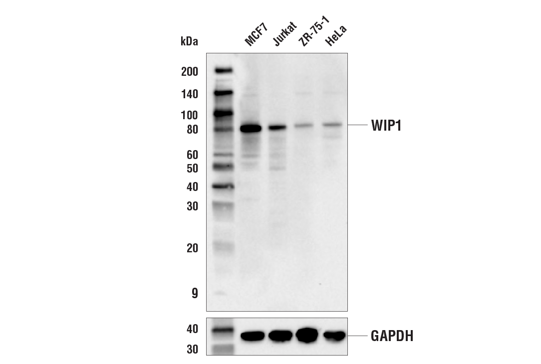
Western blot analysis of extracts from various cell lines using WIP1 (E2X1I) Rabbit mAb (upper) or GAPDH (D16H11) XP® Rabbit mAb #5174 (lower).
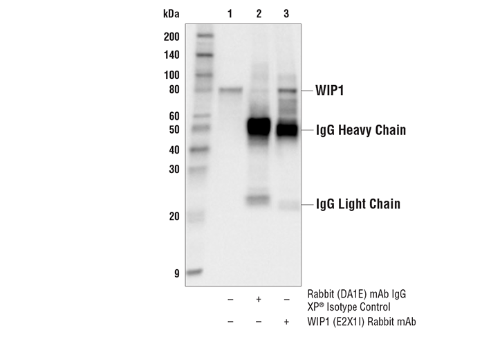
Immunoprecipitation of WIP1 protein from MCF7 cell extracts. Lane 1 is 10% input, lane 2 is Rabbit (DA1E) mAb IgG XP® Isotype Control #3900, and lane 3 is WIP1 (E2X1I) Rabbit mAb. Western blot analysis was performed using WIP1 (E2X1I) Rabbit mAb. Anti-rabbit IgG, HRP-linked Antibody #7074 was used as a secondary antibody.
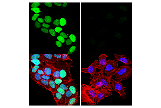
Confocal immunofluorescent analysis of MCF7 cells (left, high-expressing) or HeLa cells (right, low-expressing) using WIP1 (E2X1I) Rabbit mAb (green). Actin filaments were labeled with DyLight™ 554 Phalloidin #13054 (red). Samples were mounted in ProLong® Gold Antifade Reagent with DAPI #8961 (blue).