Revision 1
#56175
Store at -20C
Xenophagy Antibody Sampler Kit
1 Kit
(9 x 20 microliters)
877-616-CELL (2355)
877-678-TECH (8324)
3 Trask Lane | Danvers | Massachusetts | 01923 | USA
For Research Use Only. Not for Use in Diagnostic Procedures.
| Product Includes | Product # | Quantity | Mol. Wt | Isotype/Source |
|---|---|---|---|---|
| LRSAM1 (D1O5S) Rabbit mAb | 28405 | 20 µl | 84 kDa | Rabbit IgG |
| Galectin-3/LGALS3 (E7B6R) Rabbit mAb | 89572 | 20 µl | 28 kDa | Rabbit IgG |
| NDP52 (D1E4A) Rabbit mAb | 60732 | 20 µl | 52, 60 kDa | Rabbit IgG |
| TAX1BP1 (D1D5) Rabbit mAb | 5105 | 20 µl | 92 kDa | Rabbit IgG |
| Atg16L1 (D6D5) Rabbit mAb | 8089 | 20 µl | 66, 68 kDa | Rabbit IgG |
| Phospho-Atg16L1 (Ser278) (E7K6H) Rabbit mAb | 45511 | 20 µl | 68 kDa | Rabbit IgG |
| Phospho-TBK1/NAK (Ser172) (D52C2) XP® Rabbit mAb | 5483 | 20 µl | 84 kDa | Rabbit IgG |
| TBK1/NAK (E8I3G) Rabbit mAb | 38066 | 20 µl | 84 kDa | Rabbit IgG |
| LC3B (E7X4S) XP® Rabbit mAb | 43566 | 20 µl | 14, 16 kDa | Rabbit IgG |
| Anti-rabbit IgG, HRP-linked Antibody | 7074 | 100 µl | Goat |
Please visit cellsignal.com for individual component applications, species cross-reactivity, dilutions, protocols, and additional product information.
Description
The Xenophagy Antibody Sampler Kit provides an economical means of detecting selected targets involved in the process of xenophagy. The kit includes enough antibodies to perform two western blot experiments with each primary antibody.
Storage
Background
Xenophagy provides an important defense against foreign pathogens, such as bacteria and viruses, by targeting them for degradation through autophagy (1-3). Several well-known pathogens can be targeted for degradation through xenophagy, including Salmonella typhimurium, Streptococcus pyogenes, and Mycobacterium tuberculosis, and loss of core autophagy genes can result in enhanced vulnerability to these infections. Pathogens are generally targeted to LC3B or other Atg8 family members localized on autophagosome membranes. Sequestosome 1/p62-like receptors (SLRs) function as cargo receptors through ubiquitin-mediated interactions along with LIR-domain interactions with Atg8 proteins on the autophagosome. The SLR TAX1BP1 is recruited to ubiquitylated Salmonella and plays a key role in xenophagy (4). Another SLR NDP52 has also been reported to be required for xenophagy of several pathogens (5,6). LRSAM1 is an E3 ubiquitin ligase with a leucine-rich repeat recognized by microbial ligands and recruits NDP52 (7). In addition, galectins are a family of cytosolic lectins that are associated with phagophores and bacteria. Galectin-3 is recruited to bacterial membranes and is important for protection against Mycobacterium tuberculosis (8). TBK1 is a serine/threonine kinase that plays a major role in controlling autophagy by targeting substrates in the SLR family and the maturation of autophagosomes. TBK1 is phosphorylated at Ser172 within its activation loop, which is required for its kinase activity (9). Atg16L1 is a component of the Atg12-Atg5 conjugation system required for autophagosome maturation (10,11). ULK1-mediated phosphorylation of Atg16L1 at Ser278 promotes xenophagy (12).
Background References
- Reggio, A. et al. (2020) Exp Cell Res 396, 112276.
- Levine, B. (2005) Cell 120, 159-62.
- Deretic, V. et al. (2013) Nat Rev Immunol 13, 722-37.
- Tumbarello, D.A. et al. (2015) PLoS Pathog 11, e1005174.
- Verlhac, P. et al. (2015) Cell Host Microbe 17, 515-25.
- Fan, S. et al. (2020) Int J Mol Sci 21, 2008. doi: 10.3390/ijms21062008.
- Huett, A. et al. (2012) Cell Host Microbe 12, 778-90.
- Kumar, S. et al. (2017) Autophagy 13, 1086-1087.
- Kishore, N. et al. (2002) J Biol Chem 277, 13840-7.
- Fujita, N. et al. (2008) Mol Biol Cell 19, 2092-100.
- Mizushima, N. et al. (2003) J Cell Sci 116, 1679-88.
- Alsaadi, R.M. et al. (2019) EMBO Rep 20, e46885.
Trademarks and Patents
Cell Signaling Technology is a trademark of Cell Signaling Technology, Inc.
XP is a registered trademark of Cell Signaling Technology, Inc.
All other trademarks are the property of their respective owners. Visit cellsignal.com/trademarks for more information.
Limited Uses
Except as otherwise expressly agreed in a writing signed by a legally authorized representative of CST, the following terms apply to Products provided by CST, its affiliates or its distributors. Any Customer's terms and conditions that are in addition to, or different from, those contained herein, unless separately accepted in writing by a legally authorized representative of CST, are rejected and are of no force or effect.
Products are labeled with For Research Use Only or a similar labeling statement and have not been approved, cleared, or licensed by the FDA or other regulatory foreign or domestic entity, for any purpose. Customer shall not use any Product for any diagnostic or therapeutic purpose, or otherwise in any manner that conflicts with its labeling statement. Products sold or licensed by CST are provided for Customer as the end-user and solely for research and development uses. Any use of Product for diagnostic, prophylactic or therapeutic purposes, or any purchase of Product for resale (alone or as a component) or other commercial purpose, requires a separate license from CST. Customer shall (a) not sell, license, loan, donate or otherwise transfer or make available any Product to any third party, whether alone or in combination with other materials, or use the Products to manufacture any commercial products, (b) not copy, modify, reverse engineer, decompile, disassemble or otherwise attempt to discover the underlying structure or technology of the Products, or use the Products for the purpose of developing any products or services that would compete with CST products or services, (c) not alter or remove from the Products any trademarks, trade names, logos, patent or copyright notices or markings, (d) use the Products solely in accordance with CST Product Terms of Sale and any applicable documentation, and (e) comply with any license, terms of service or similar agreement with respect to any third party products or services used by Customer in connection with the Products.
Revision 1
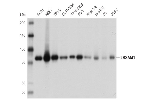
Western blot analysis of extracts from various cell lines using LRSAM1 (D1O5S) Rabbit mAb.
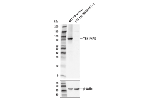
Western blot analysis of extracts from HCT 116 cells, either wild-type (+/+) or TBK1/NAK knockout (-/-), using TBK1/NAK (E8I3G) Rabbit mAb (upper) or β-Actin (D6A8) Rabbit mAb #8457 (lower).
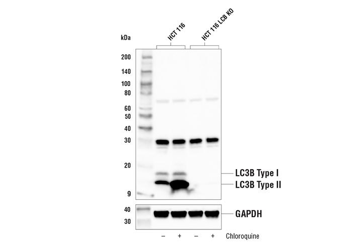
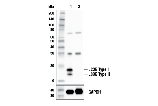
Western blot analysis of extracts from HCT 116 and HCT 116 LC3B knockout cells, untreated (-) or treated with Chloroquine #14774 (50 μM, 18 hr) using LC3B (E7X4S) XP® Rabbit mAb #43566 (upper) or GAPDH (D16H11) XP® Rabbit mAb #5174 (lower). The absence of signal in the HCT 116 knockout cells confirms the specificity of the antibody for LC3B.
Revision 1
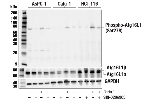
Western blot analysis of extracts from AsPC-1, Calu-1, and HCT 116 cells, untreated (-) or treated with Torin 1 #14379 (250 nM, 2 hr; +) and/or the ULK1 inhibitor SBI-0206965 #29089 (50 μM, 2 hr; +), using Phospho-Atg16L1 (Ser278) (E7K6H) Rabbit mAb (upper), Atg16L1 (D6D5) Rabbit mAb #8089 (middle), or GAPDH (D16H11) XP® Rabbit mAb #5174 (lower).
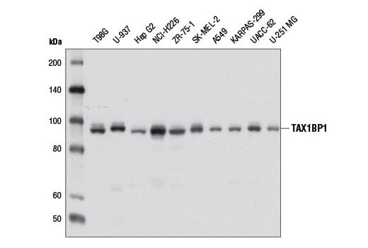
Western blot analysis of extracts from various cell lines using TAX1BP1 (D1D5) Rabbit mAb.
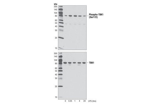
Western blot analysis of extracts from THP-1 cells differentiated with TPA #4174 (80 nM, overnight) followed by treatment with LPS (1 μg/ml), up to 24h, using Phospho-TBK1/NAK (Ser172) (D52C2) XP® Rabbit mAb (upper), or total TBK1/NAK (D1B4) Rabbit mAb #3504 (lower).
Revision 1
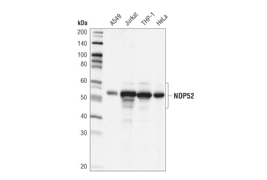
Western blot analysis of extracts from various cell lines using NDP52 (D1E4A) Rabbit mAb.
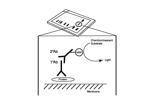
After the primary antibody is bound to the target protein, a complex with HRP-linked secondary antibody is formed. The LumiGLO® is added and emits light during enzyme catalyzed decomposition.
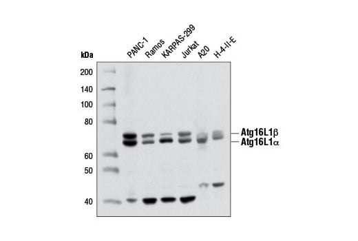
Western blot analysis of extracts from various cell lines using Atg16L1 (D6D5) Rabbit mAb.
Revision 1
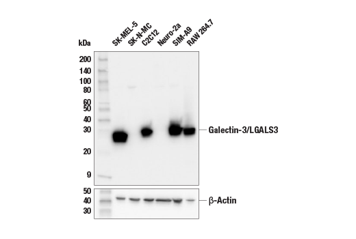
Western blot analysis of extracts from various cell lines using Galectin-3/LGALS3 (E7B6R) Rabbit mAb (upper) and β-Actin (D6A8) Rabbit mAb #8457 (lower).
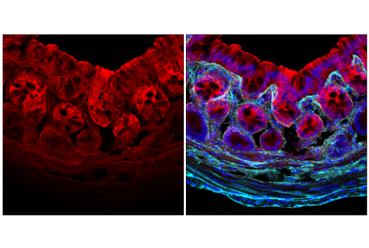
Confocal immunofluorescent analysis of fixed frozen mouse colon labeled with Galectin-3/LGALS3 (E7B6R) Rabbit mAb (left, red). Free secondary binding sites were then blocked with Rabbit (DA1E) mAb IgG XP® Isotype Control #3900 prior to colabeling with COL1A1 (E8F4L) XP® Rabbit mAb (Alexa Fluor® 647 Conjugate) #72827 (right, cyan pseudocolor) and DAPI #4083 (right, blue).
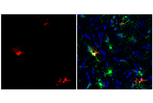
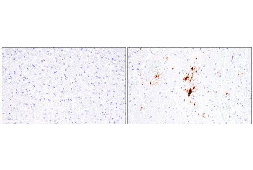
Confocal immunofluorescent analysis of fixed frozen mouse subicular cortex from an amyloid mouse model of Alzheimer's Disease labeled with Galectin-3/LGALS3 (E7B6R) Rabbit mAb (left, red). Free secondary binding sites were then blocked with Rabbit (DA1E) mAb IgG XP® Isotype Control #3900 prior to colabeling with Iba1/AIF-1 (E4O4W) XP® Rabbit mAb (Alexa Fluor® 488 Conjugate) #20825 (right, green) and DAPI #4083 (right, blue).
Revision 1
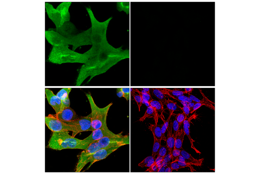
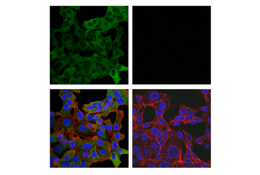
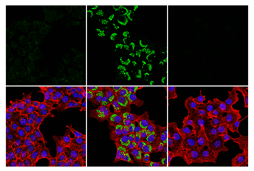
Confocal immunofluorescent analysis of SK-MEL-5 cells (left, positive) and SK-N-MC cells (right, negative) using Galectin-3/LGALS3 (E7B6R) Rabbit mAb (green), DyLight™ 554 Phalloidin #13054 (red), and DAPI #4083 (blue).
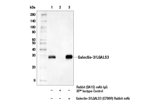
Immunoprecipitation of Galectin-3/LGALS3 protein from C2C12 cell extracts. Lane 1 is 10% input, lane 2 is Rabbit (DA1E) mAb IgG XP® Isotype Control #3900, and lane 3 is Galectin-3/LGALS3 (E7B6R) Rabbit mAb. Western blot analysis was performed using Galectin-3/LGALS3 (E7B6R) Rabbit mAb. Mouse Anti-rabbit IgG (Conformation Specific) (L27A9) mAb (HRP Conjugate) #5127 was used as a secondary antibody.
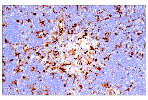
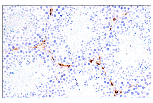
Immunohistochemical analysis of paraffin-embedded mouse thymus using Galectin-3/LGALS3 (E7B6R) Rabbit mAb.
Revision 1
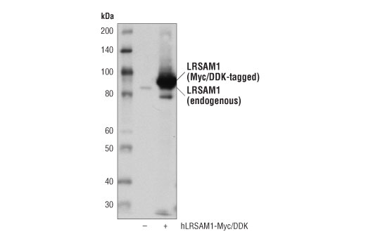
Western blot analysis of extracts from 293T cells, mock transfected (-) or transfected with a construct expressing Myc/DDK-tagged full-length human LRSAM1 protein (hLRSAM1-Myc/DDK; +), using LRSAM1 (D1O5S) Rabbit mAb.
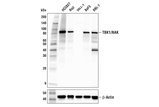
Western blot analysis of extracts from various cell lines using TBK1/NAK (E8I3G) Rabbit mAb (upper) or β-Actin (D6A8) Rabbit mAb #8457 (lower).
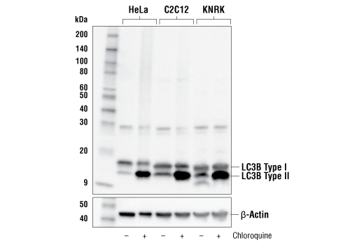
Western blot analysis of extracts from HeLa, C2C12, and KNRK cells, untreated (-) or treated with Chloroquine #14774 (50 μM, 18 hr; +), using LC3B (E7X4S) XP® Rabbit mAb (upper) or β-Actin (D6A8) Rabbit mAb #8457 (lower).
Revision 1
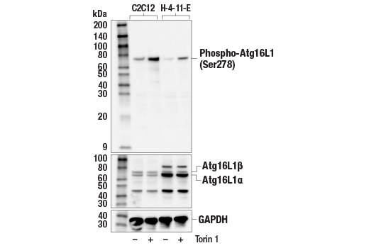
Western blot analysis of extracts from C2C12 and H-4-II-E cells, untreated (-) or treated with Torin 1 #14379 (250 nM, 2 hr; +), using Phospho-Atg16L1 (Ser278) (E7K6H) Rabbit mAb (upper), Atg16L1 (D6D5) Rabbit mAb #8089 (middle), or GAPDH (D16H11) XP® Rabbit mAb #5174 (lower).
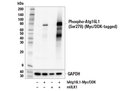
Western blot analysis of extracts from 293T cells, mock transfected (-) or transfected with constructs expressing Myc/DDK-tagged full-length human Atg16L1 protein (hAtg16L1-Myc/DDK; +) and/or mouse ULK1 protein (mULK1; +), using Phospho-Atg16L1 (Ser278) (E7K6H) Rabbit mAb (upper) or GAPDH (D16H11) XP® Rabbit mAb #5174 (lower).
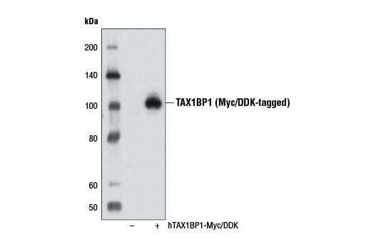
Western blot analysis of extracts from 293T cells, either mock transfected (-) or transfected with a Myc/DDK-tagged cDNA expression construct encoding the full-length isoform 1 of human TAX1BP1 (hTAX1BP1-Myc/DDK, +), using TAX1BP1 (D1D5) Rabbit mAb.
Revision 1
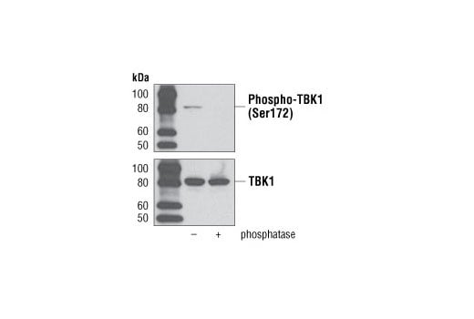
Western blot analysis of extracts from THP-1 cells differentiated with TPA #4174 (80 nM, overnight) followed by treatment with LPS (1 μg/ml, 1 hour), with or without phosphatase treatment using Phospho-TBK1/NAK (Ser172) (D52C2) XP® Rabbit mAb (upper), or total TBK1/NAK (D1B4) Rabbit mAb #3504 (lower).
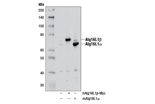
Western blot analysis of extracts from 293T cells, mock transfected (-) or transfected with either a myc-tagged human Atg16L1β construct (hAtg16L1β-Myc; +) or a mouse Atg16L1α construct (mAtg16L1α; +), using Atg16L1 (D6D5) Rabbit mAb. The myc-tagged human Atg16L1β construct was kindly provided by Dr. Qing Zhong, University of California Berkeley.
Immunohistochemical analysis of paraffin-embedded wild-type mouse brain (left) or brain from an amyloid mouse model of Alzheimer's Disease (right) using Galectin-3/LGALS3 (E7B6R) Rabbit mAb.
Revision 1
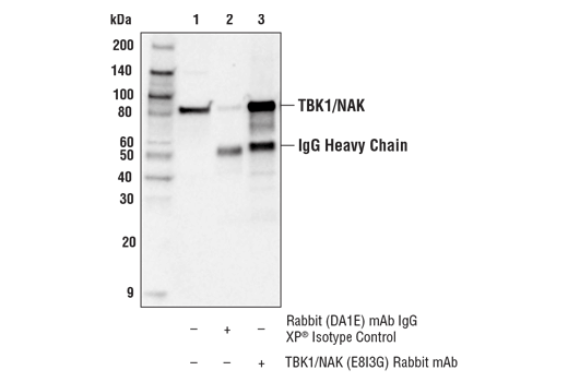
Immunoprecipitation of TBK1/NAK from Raji cell extracts. Lane 1 is 10% input, lane 2 is Rabbit (DA1E) mAb IgG XP® Isotype Control #3900, and lane 3 is TBK1/NAK (E8I3G) Rabbit mAb. Western blot analysis was performed using TBK1/NAK (E8I3G) Rabbit mAb.
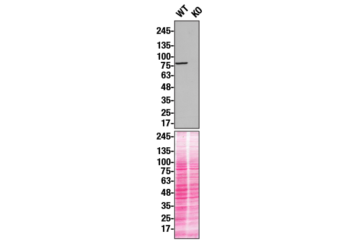
Western blot analysis of U20S extracts from WT (left) or TBK1 KO (right) using TBK1/NAK (E8I3G) Rabbit mAb. Membranes stained with Ponceau S for total protein normalization (lower). These data were provided by YCharOS Inc., an open science company with the mission of characterizing commercially available antibodies, as a companion to validation data generated by CST scientists.
Western blot analysis of extracts from control HeLa cells (lane 1) or LC3B knockout HeLa cells (lane 2) using LC3B (E7X4S) XP® Rabbit mAb (upper) or GAPDH (D16H11) XP® Rabbit mAb #5174 (lower). The absence of signal in the LC3B knockout HeLa cells confirms specificity of the antibody for LC3B.
Revision 1
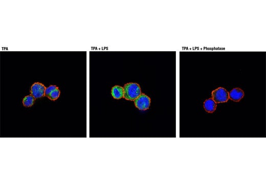
Confocal immunofluorescent analysis of THP-1 cells differentiated with TPA #4174 (80nM, overnight) (left), followed by treatment with LPS (1μg/ml, 1 hour) (center) or LPS with λ phosphatase treatment (right) using Phospho-TBK1/NAK (Ser172) (D52C2) XP® Rabbit mAb (green). Actin filaments were labeled with DY-554 Phalloidin (red). Blue pseudocolor = DRAQ5® #4084 (fluorescent DNA dye).
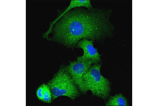
Confocal immunofluorescent analysis of EBSS-starved PANC-1 cells using Atg16L1 (D6D5) Rabbit mAb (green). Blue pseudocolor = DRAQ5® #4084 (fluorescent DNA dye).
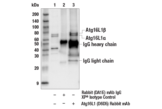
Immunoprecipitation of Atg16L1 from Jurkat cell extracts. Lane 1 is 10% input, lane 2 is precipitated with Rabbit (DA1E) mAb IgG XP® Isotype Control #3900, and lane 3 is Atg16L1 (D6D5) Rabbit mAb, #8089. Western blot was performed using Atg16L1 (D6D5) Rabbit mAb.
Revision 1
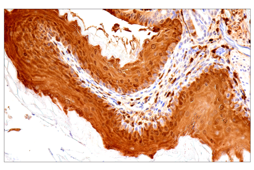
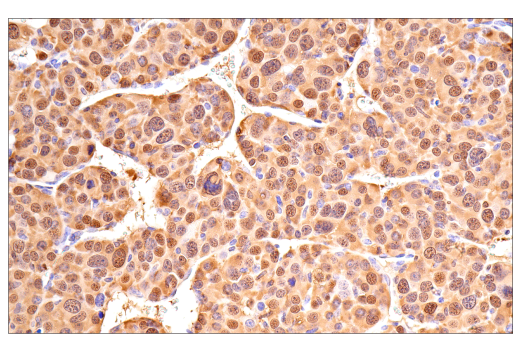
Immunohistochemical analysis of paraffin-embedded mouse spleen using Galectin-3/LGALS3 (E7B6R) Rabbit mAb.
Confocal immunofluorescent analysis of HCT 116 cells, wild-type (left, positive) or TBK1/NAK knockout (right, negative), using TBK1/NAK (E8I3G) Rabbit mAb (green). Actin filaments were labeled with Alexa Fluor® 555 Phalloidin #8953 (red). Samples were mounted in ProLong® Gold Antifade Reagent with DAPI #8961 (blue).
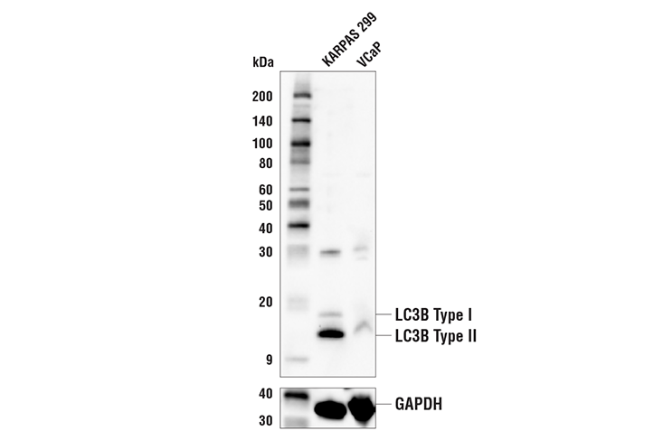
Western blot analysis from KARPAS 299 and VCap cells using LC3B (E7X4S) XP® Rabbit mAb #43566 (upper) or GAPDH (D16H11) XP® Rabbit mAb #5174 (lower). The low signal of LC3B in VCap cells is predicted from RNAseq data and confirms the specificity of the antibody for LC3B.
Revision 1
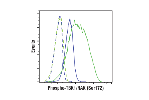
Flow cytometric analysis of THP-1 cells differentiated with TPA (80nM, 4 days) #9905, untreated (blue) or treated with LPS (1 ng/mL, 1 hr; green) #14011 using Phospho-TBK1/NAK (Ser172) (D52C2) XP® Rabbit mAb (solid lines) or concentration-matched Rabbit (DA1E) mAb IgG XP® Isotype Control #3900 (dashed lines). Anti-rabbit IgG (H+L), F(ab')2 Fragment (Alexa Fluor® 488 Conjugate) #4412 was used as a secondary antibody.
Immunohistochemical analysis of paraffin-embedded mouse testis using Galectin-3/LGALS3 (E7B6R) Rabbit mAb.
Immunohistochemical analysis of paraffin-embedded mouse forestomach using Galectin-3/LGALS3 (E7B6R) Rabbit mAb.
Revision 1
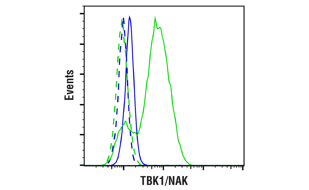
Flow cytometric analysis of TALL-1 cells (blue, negative) and Raji cells (green, positive) using TBK1/NAK (E8I3G) Rabbit mAb (solid lines) or concentration-matched Rabbit (DA1E) mAb IgG XP® Isotype Control #3900 (dashed lines). Anti-rabbit IgG (H+L), F(ab')2 Fragment (Alexa Fluor® 488 Conjugate) #4412 was used as a secondary antibody.
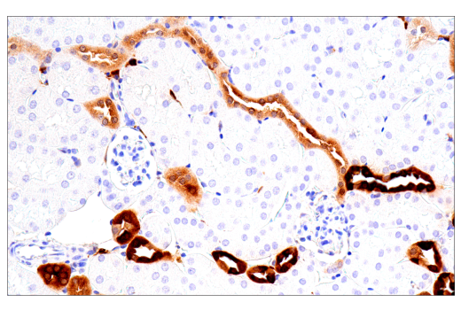
Immunohistochemical analysis of paraffin-embedded mouse kidney using Galectin-3/LGALS3 (E7B6R) Rabbit mAb.
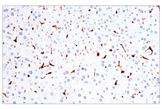
Immunohistochemical analysis of paraffin-embedded mouse liver using Galectin-3/LGALS3 (E7B6R) Rabbit mAb.
Revision 1
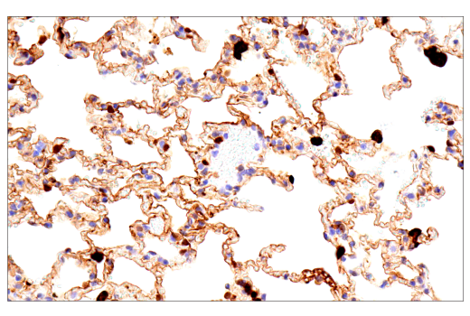
Immunohistochemical analysis of paraffin-embedded mouse lung using Galectin-3/LGALS3 (E7B6R) Rabbit mAb.
Immunohistochemical analysis of paraffin-embedded mouse ovary using Galectin-3/LGALS3 (E7B6R) Rabbit mAb.
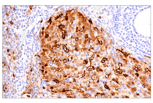
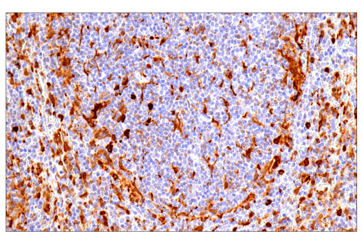
Immunohistochemical analysis of paraffin-embedded A20 syngeneic tumor using Galectin-3/LGALS3 (E7B6R) Rabbit mAb.
Revision 1
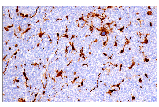
Immunohistochemical analysis of paraffin-embedded GL261 syngeneic tumor using Galectin-3/LGALS3 (E7B6R) Rabbit mAb.
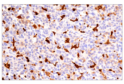
Immunohistochemical analysis of paraffin-embedded Renca syngeneic tumor using Galectin-3/LGALS3 (E7B6R) Rabbit mAb.
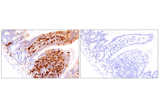
Immunohistochemical analysis of paraffin-embedded mouse small intestine using Galectin-3/LGALS3 (E7B6R) Rabbit mAb (left) compared to concentration-matched Rabbit (DA1E) mAb IgG XP® Isotype Control #3900 (right).
Revision 1
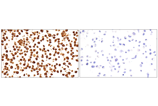
Immunohistochemical analysis of paraffin-embedded RAW 264.7 cell pellet (left, positive) or Neuro-2a cell pellet (right, negative) using Galectin-3/LGALS3 (E7B6R) Rabbit mAb.
Confocal immunofluorescent analysis of HCT 116 cells either untreated (left) or treated with Chloroquine #14774 (50 µM, overnight) (center) or LC3B HCT 116 knockout cells treated with Chloroquine #14774 (50 µM, overnight) (right) using LC3B (E7X4S) XP® Rabbit mAb (green). Actin filaments were labeled with β-Actin (8H10D10) Mouse mAb (red) and nuclei were labeled with DAPI #4083 (blue).
Confocal immunofluorescent analysis of MIA PaCa-2 cells untreated (left), nutrient-starved with EBSS (2 hr; middle), or treated with Chloroquine #14774 (50 µM, 24 hr; right) using LC3B (E7X4S) XP® Rabbit mAb (green) and β-Actin (8H10D10) Mouse mAb #3700 (red). Samples were mounted in ProLong® Gold Antifade Reagent with DAPI #8961 (blue).
Revision 1
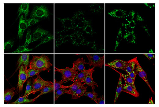
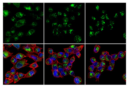
Confocal immunofluorescent analysis of C2C12 cells untreated (left), nutrient-starved with EBSS (2 hr; middle), or treated with Chloroquine #14774 (50 µM, 24 hr; right) using LC3B (E7X4S) XP® Rabbit mAb (green) and β-Actin (8H10D10) Mouse mAb #3700 (red). Samples were mounted in ProLong® Gold Antifade Reagent with DAPI #8961 (blue).