Revision 1
#53360
Store at -20C
Pan-Keratin (Type I) (E6S1S) & CO-0072-750 SignalStar™ Oligo-Antibody Pair
1 Kit
(10 slides)
UniProt ID:
#P02535, #P05784, #Q61781, #Q61414, #Q9Z2K1, #Q9QWL7, #P19001
Entrez-Gene Id:
#16661, #16668, #16664, #16665, #16666, #16667, #16669
877-616-CELL (2355)
877-678-TECH (8324)
3 Trask Lane | Danvers | Massachusetts | 01923 | USA
For Research Use Only. Not for Use in Diagnostic Procedures.
| Product Includes | Product # | Volume | Reactivity | Isotype |
|---|---|---|---|---|
| Pan-Keratin (Type I) (E6S1S) Rabbit mAb (SignalStar™ Conjugate 0072) | 74839 | 50 µl | H M | Rabbit IgG |
| Complementary Oligo (CO-0072-750) | 66642 | 22 µl |
Storage
Description
SignalStar multiplex immunohistochemistry (IHC) is an advanced technology for labeling multiple proteins simultaneously in tissue samples using specific primary antibodies and fluorescent detection reagents. This technology offers accuracy and reliability in visualizing and analyzing protein expression while maintaining spatial context and tissue architecture.
SignalStar Oligo-Antibody Pairs are compatible with the SignalStar Multiplex IHC Buffer Kits for use in fluorescent multiplex imaging experiments. This product includes the oligo-conjugated antibodies and complementary oligos required for labeling your target protein on up to 10 slides. SignalStar Multiplex IHC Buffer Kits are required to amplify and image the target signal. Multiple oligo-antibody pairs can be conveniently combined into a multiplex panel using the SignalStar Multiplex IHC Panel Builder. SignalStar Multiplex IHC Kits & Reagents are not compatible with all of Cell Signaling Technology® products and protocols that are recommended for use in immunohistochemical assays.
Specificity/Sensitivity
Pan-Keratin (Type I) (E6S1S) Rabbit mAb (SignalStar™ Conjugate 0072) recognizes endogenous levels of total type I keratin protein. The antibody was validated to detect overexpressed keratin 10, 17, and 18. By homology, it is also predicted to detect keratin 13, 14, 15, 16, and 19. It does not detect type II keratins including keratin 1, 5, 6a, 7, and 8. Non-specific staining was observed in Leydig cells of the testis by immunohistochemistry.
Source / Purification
Monoclonal antibody is produced by immunizing animals with a mix of synthetic peptides corresponding to residues highly conserved among type I keratins.
Background
Keratins (cytokeratins) are intermediate filament proteins that are mainly expressed in epithelial cells. Keratin heterodimers composed of an acidic keratin (or type I keratin, keratins K9-K28) and a basic keratin (or type II keratin, keratins K1-K8 and K71-K80) assemble to form filaments. Keratin isoforms demonstrate tissue- and differentiation-specific profiles that make them useful as research and clinical biomarkers (1,2).
Dysregulation/mutations in keratin genes can lead to a variety of disorders affecting the skin, hair, nails, and other epithelial tissues (3). While expression of keratins can be variable, immunohistochemical staining of keratins is widely used to help in the identification and classification of epithelial tumors, and may also provide prognostic information.
Keratins 8 and 18 (K8/K18) are expressed in simple epithelia of normal tissue, as well as in adenocarcinomas of the breast, lung, ovary, and gastrointestinal tract. Keratin 17 is expressed in basal keratinocytes of stratified epithelia, hair follicles, and sebaceous glands. Onset of keratin 17 expression coincides with the definition of major epithelial lineages during skin development (4). Keratin 14 (K14) is expressed in basal cells of stratified epithelia, and in basal-like subtypes of breast cancer and squamous cell carcinomas. Keratin 19 (K19) is expressed in glandular epithelia, including the liver, gallbladder, and pancreas, as well as in adenocarcinomas of the breast, thyroid, and bile duct. Keratin 20 (K20) is expressed in gastrointestinal epithelium, urothelium, and Merkel cells in the skin, as well as in colorectal carcinomas and some urothelial carcinomas. Keratin 5/6 (K5/6) is expressed in basal cells of stratified epithelia, including the skin, prostate, and breast, as well as in basal-like breast cancers, squamous cell carcinomas, and some lung carcinomas. Keratin 7 (K7) is expressed in glandular epithelia, such as those in the lung, breast, and female reproductive tract, as well as in adenocarcinomas of the lung, breast, and ovary (5,6).
Keratins, particularly K8, K18, and K19, serve as biomarkers for identification of circulating tumor cells (CTCs) (5).
Post-translational modifications, including phosphorylation, acetylation, ubiquitylation, sumoylation, glycosylation, and transamidation, have been shown to affect the functions of keratins in normal and disease states (6). Understanding the molecular mechanisms underlying these PTMs may provide insights into cancer pathogenesis.
Background References
- Chang, L. and Goldman, R.D. (2004) Nat Rev Mol Cell Biol 5, 601-13.
- Schweizer, J. et al. (2006) J Cell Biol 174, 169-74.
- Sarma, A. (2022) Int J Biol Macromol 219, 395-413.
- McGowan, K.M. and Coulombe, P.A. (1998) J Cell Biol 143, 469-86.
- Werner, S. et al. (2020) Mol Aspects Med 72, 100817.
- Dmello, C. et al. (2019) J Biosci 44, 33.
Species Reactivity
Species reactivity is determined by testing in at least one approved application (e.g., western blot).
Applications Key
StarBond: SignalStar™ Leica Bond
Cross-Reactivity Key
H: Human M: Mouse R: Rat Hm: Hamster Mk: Monkey Vir: Virus Mi: Mink C: Chicken Dm: D. melanogaster X: Xenopus Z: Zebrafish B: Bovine Dg: Dog Pg: Pig Sc: S. cerevisiae Ce: C. elegans Hr: Horse GP: Guinea Pig Rab: Rabbit G: Goat All: All Species Expected
Trademarks and Patents
Cell Signaling Technology is a trademark of Cell Signaling Technology, Inc.
SignalStar is a trademark of Cell Signaling Technology, Inc.
U.S. Patent No. 10,781,477, foreign equivalents, and child patents deriving therefrom.
All other trademarks are the property of their respective owners. Visit cellsignal.com/trademarks for more information.
Limited Uses
Except as otherwise expressly agreed in a writing signed by a legally authorized representative of CST, the following terms apply to Products provided by CST, its affiliates or its distributors. Any Customer's terms and conditions that are in addition to, or different from, those contained herein, unless separately accepted in writing by a legally authorized representative of CST, are rejected and are of no force or effect.
Products are labeled with For Research Use Only or a similar labeling statement and have not been approved, cleared, or licensed by the FDA or other regulatory foreign or domestic entity, for any purpose. Customer shall not use any Product for any diagnostic or therapeutic purpose, or otherwise in any manner that conflicts with its labeling statement. Products sold or licensed by CST are provided for Customer as the end-user and solely for research and development uses. Any use of Product for diagnostic, prophylactic or therapeutic purposes, or any purchase of Product for resale (alone or as a component) or other commercial purpose, requires a separate license from CST. Customer shall (a) not sell, license, loan, donate or otherwise transfer or make available any Product to any third party, whether alone or in combination with other materials, or use the Products to manufacture any commercial products, (b) not copy, modify, reverse engineer, decompile, disassemble or otherwise attempt to discover the underlying structure or technology of the Products, or use the Products for the purpose of developing any products or services that would compete with CST products or services, (c) not alter or remove from the Products any trademarks, trade names, logos, patent or copyright notices or markings, (d) use the Products solely in accordance with CST Product Terms of Sale and any applicable documentation, and (e) comply with any license, terms of service or similar agreement with respect to any third party products or services used by Customer in connection with the Products.
Revision 1
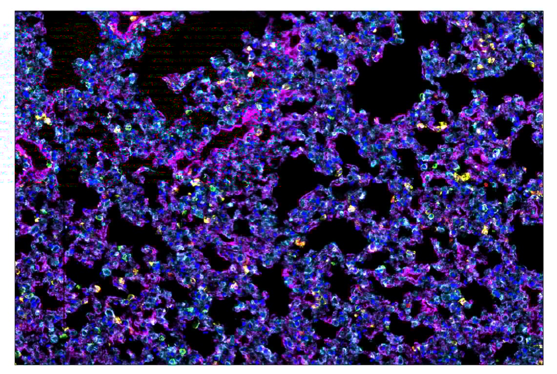
SignalStar™ multiplex immunohistochemical analysis of paraffin-embedded mouse lung using CD11b/ITGAM (E4K8C) & CO-0083-594 SignalStar™ Oligo-Antibody Pair #84898 (yellow), CD39/NTPDase 1 (E2X6B) & CO-0076-647 SignalStar™ Oligo-Antibody Pair #88633 (magenta), Pan-Keratin (Type I) (E6S1S) & CO-0072-750 SignalStar™ Oligo-Antibody Pair #53360 (cyan), CD3ε (E4T1B) & CO-0048-488 SignalStar™ Oligo-Antibody Pair #92858 (green), F4/80 (D2S9R) & CO-0042-750 SignalStar™ Oligo-Antibody Pair #51924 (red), and DAPI #4083 (blue). All fluorophores have been assigned a pseudocolor, as indicated. Staining was performed on the BOND RX autostainer by Leica Biosystems.
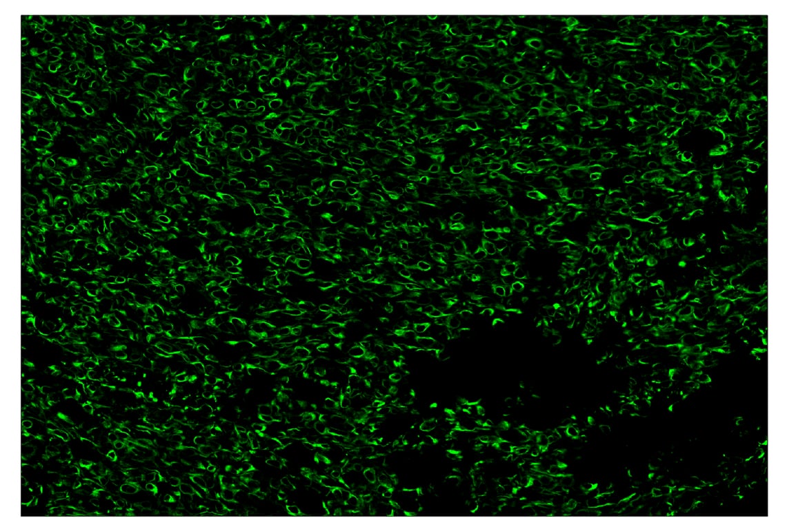
SignalStar™ multiplex immunohistochemical analysis of paraffin-embedded LL/2 syngeneic tumor using Pan-Keratin (Type I) (E6S1S) & CO-0072-488 SignalStar™ Oligo-Antibody Pair #95530 (green). All fluorophores have been assigned a pseudocolor, as indicated. Staining was performed on the BOND RX autostainer by Leica Biosystems.
SignalStar™ multiplex immunohistochemical analysis of paraffin-embedded mouse thymus using Pan-Keratin (Type I) (E6S1S) & CO-0072-594 SignalStar™ Oligo-Antibody Pair #30754 (yellow). All fluorophores have been assigned a pseudocolor, as indicated. Staining was performed on the BOND RX autostainer by Leica Biosystems.
Revision 1
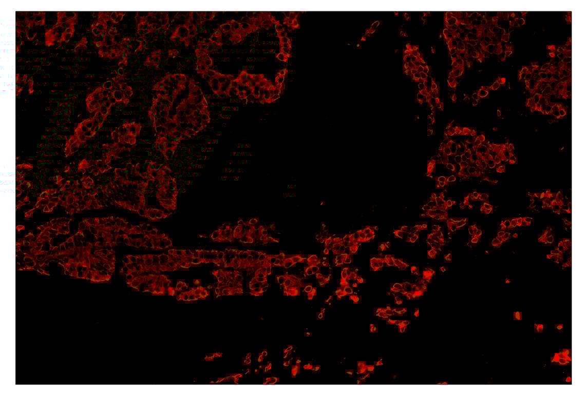
SignalStar™ multiplex immunohistochemical analysis of paraffin-embedded human non-small cell lung carcinoma using Pan-Keratin (Type I) (E6S1S) & CO-0072-647 SignalStar™ Oligo-Antibody Pair #77030 (red). All fluorophores have been assigned a pseudocolor, as indicated. Staining was performed on the BOND RX autostainer by Leica Biosystems.
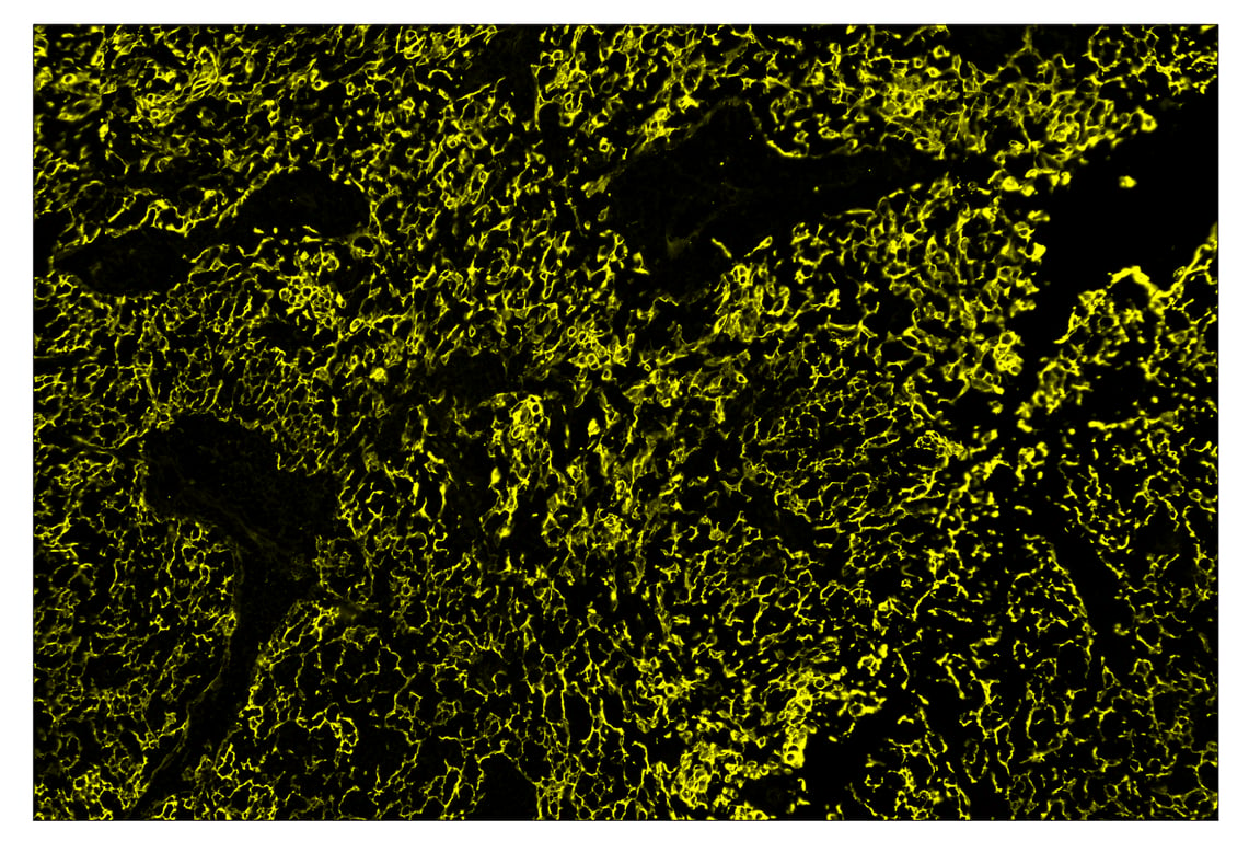
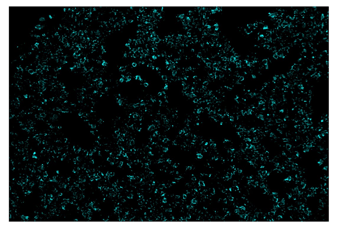
SignalStar™ multiplex immunohistochemical analysis of paraffin-embedded mouse lung using Pan-Keratin (Type I) (E6S1S) & CO-0072-750 SignalStar™ Oligo-Antibody Pair #53360 (cyan). All fluorophores have been assigned a pseudocolor, as indicated. Staining was performed on the BOND RX autostainer by Leica Biosystems.
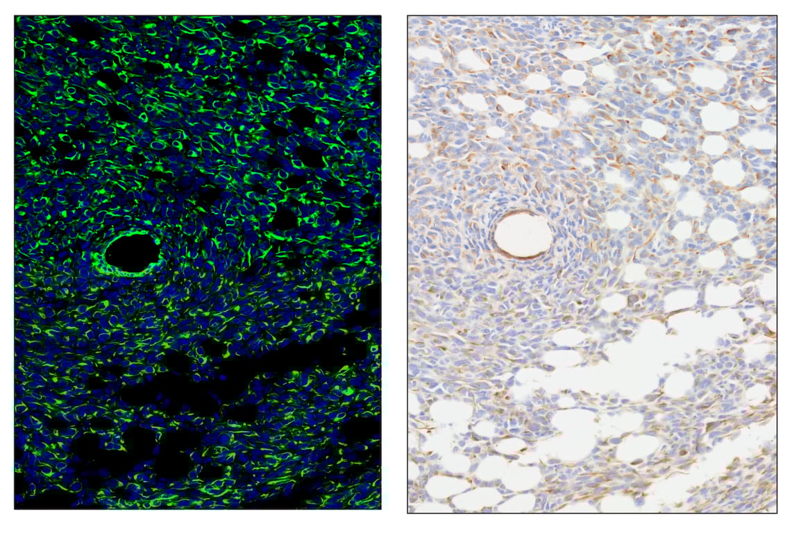
SignalStar™ multiplex immunohistochemical analysis of paraffin-embedded LL/2 syngeneic tumor using Pan-Keratin (Type I) (E6S1S) & CO-0072-488 SignalStar™ Oligo-Antibody Pair #95530 (left, yellow) and DAPI #4083 (left, blue) compared to chromogenic immunohistochemical analysis of a serial section of paraffin-embedded LL/2 syngeneic tumor using Pan-Keratin (Type I) (E6S1S) Rabbit mAb #83957 (right). All fluorophores have been assigned a pseudocolor, as indicated. Staining was performed on the BOND RX autostainer by Leica Biosystems.
Revision 1
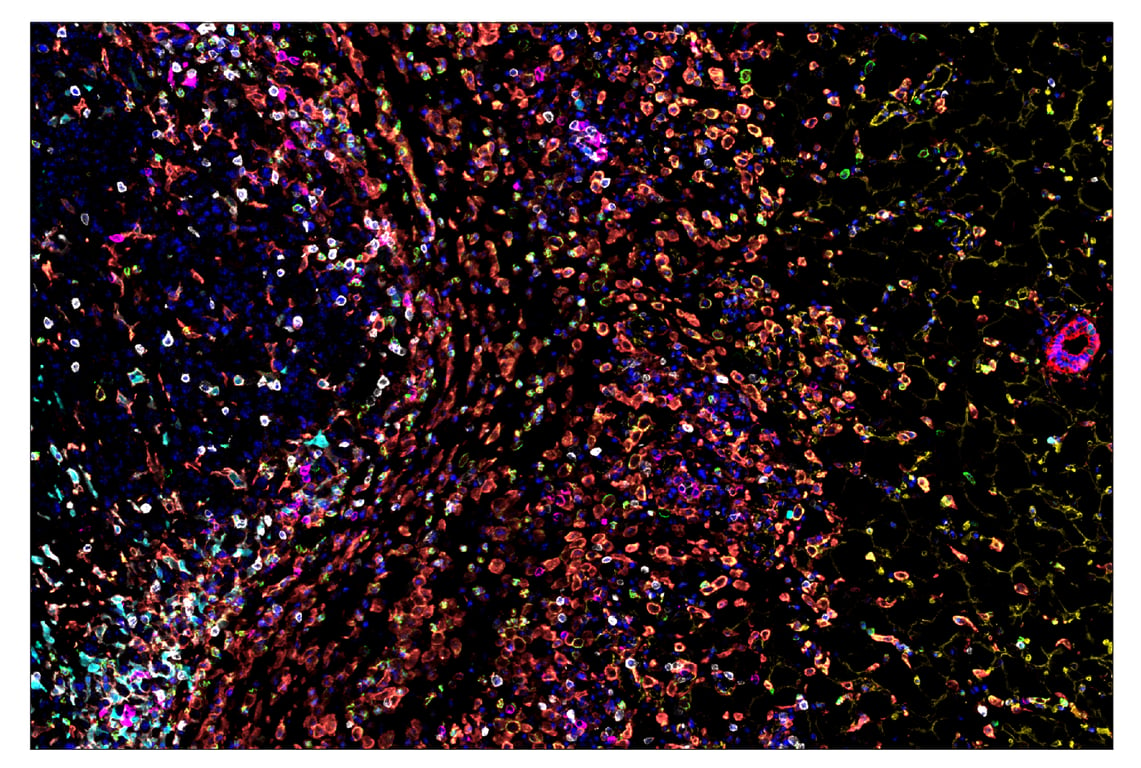
SignalStar™ multiplex immunohistochemical analysis of paraffin-embedded GL261 syngeneic tumor using CD11b/ITGAM (E4K8C) & CO-0083-488 SignalStar™ Oligo-Antibody Pair #39172 (green), CD36 (E8B7S) & CO-0089-594 SignalStar™ Oligo-Antibody Pair #23418 (yellow), Pan-Keratin (Type I) (E6S1S) & CO-0072-647 SignalStar™ Oligo-Antibody Pair #77030 (red), IL-2Rα/CD25 (E9W2J) & CO-0074-750 SignalStar™ Oligo-Antibody Pair #71915 (magenta), CD8α (D4W2Z) & CO-0040-488 SignalStar™ Oligo-Antibody Pair #17707 (white), Arginase-1 (D4E3M) & CO-0075-594 SignalStar™ Oligo-Antibody Pair #66757 (cyan), F4/80 (D2S9R) & CO-0042-750 SignalStar™ Oligo-Antibody Pair #51924 (orange), and DAPI #4083 (blue). All fluorophores have been assigned a pseudocolor, as indicated. Staining was performed on the BOND RX autostainer by Leica Biosystems.
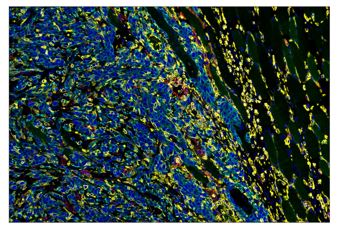
SignalStar™ multiplex immunohistochemical analysis of paraffin-embedded 4T1 syngeneic mammary tumor using CD36 (E8B7S) & CO-0089-488 SignalStar™ Oligo-Antibody Pair #95886 (green), CD11b/ITGAM (E4K8C) & CO-0083-594 SignalStar™ Oligo-Antibody Pair #84898 (yellow), CD39/NTPDase 1 (E2X6B) & CO-0076-647 SignalStar™ Oligo-Antibody Pair #88633 (red), Pan-Keratin (Type I) (E6S1S) & CO-0072-750 SignalStar™ Oligo-Antibody Pair #53360 (cyan), and DAPI #4083 (blue). All fluorophores have been assigned a pseudocolor, as indicated.