Revision 1

#90033

Store at -20C

MHC Class II (LGII-612.14) & CO-0105-594 SignalStar Oligo-Antibody Pair

1 Kit

(10 slides)

UniProt ID:
#P01911, #P04440

Entrez-Gene Id:
#3123, #3115

CST Logo
Orders:

877-616-CELL (2355)

orders@cellsignal.com

Support:

877-678-TECH (8324)

3 Trask Lane | Danvers | Massachusetts | 01923 | USA

For Research Use Only. Not for Use in Diagnostic Procedures.

Product Includes Product # Volume Reactivity Isotype
MHC Class II (LGII-612.14) Mouse mAb (SignalStar™ Conjugate 0105)7910150 µlHMouse IgG1
Complementary Oligo (CO-0105-594)4845022 µl

Storage

SignalStar conjugates are supplied in PBS (pH 7.2), less than 0.1% sodium azide, 2 mM EDTA, 0.05% Triton X-100, 2 mg/mL BSA, and 50% glycerol. Complementary oligos are supplied in nuclease-free water. Store at -20°C. Do not aliquot the antibody. All components in this kit are stable for at least 12 months when stored at the recommended temperature.

Description

SignalStar multiplex immunohistochemistry (IHC) is an advanced technology for labeling multiple proteins simultaneously in tissue samples using specific primary antibodies and fluorescent detection reagents. This technology offers accuracy and reliability in visualizing and analyzing protein expression while maintaining spatial context and tissue architecture.

SignalStar Oligo-Antibody Pairs are compatible with the SignalStar Multiplex IHC Buffer Kits for use in fluorescent multiplex imaging experiments. This product includes the oligo-conjugated antibodies and complementary oligos required for labeling your target protein on up to 10 slides. SignalStar Multiplex IHC Buffer Kits are required to amplify and image the target signal. Multiple oligo-antibody pairs can be conveniently combined into a multiplex panel using the SignalStar Multiplex IHC Panel Builder. SignalStar Multiplex IHC Kits & Reagents are not compatible with all of Cell Signaling Technology® products and protocols that are recommended for use in immunohistochemical assays.

Specificity/Sensitivity

MHC Class II (LGII-612.14) Mouse mAb (SignalStar™ Conjugate 0105) exhibits strong reactivity with HLA-DRB and weak reactivity with HLA-DPB in cell lines transfected with constructs expressing Myc/DDK-tagged HLA-DRB and HLA-DPB, respectively. Reactivity is not observed with HLA-DMB, HLA-DOB, and HLA-DQB in cell lines transfected with constructs expressing Myc/DDK-tagged HLA-DMB, HLA-DOB, and HLA-DQB.

Source / Purification

Monoclonal antibody is produced by immunizing animals with cultured human B lymphoid cells treated with IFN-gamma.

Background

Major histocompatibility complex class II (MHC class II) molecules are heterodimeric, transmembrane glycoproteins expressed on the surface of antigen-presenting cells, such as macrophages, dendritic cells, and B cells. Expression can also be induced on other cell types through interferon-γ signaling (1). Prior to being displayed on the cell membrane, MHC class II molecules are loaded with exogenous peptide antigens approximately 15-24 amino acids in length that were derived from endocytosed extracellular proteins digested in the lysosome (2). Antigen-presentation through MHC class II is required for T cell activation during the immune response to extracellular pathogens (2). In humans, the MHC class II protein complex is encoded by the human leukocyte antigen gene complex (HLA). HLAs corresponding to MHC class II are HLA-DP, HLA-DM, HLA-DOA, HLA-DOB, HLA-DQ, and HLA-DR (3).

Species Reactivity

Species reactivity is determined by testing in at least one approved application (e.g., western blot).

Trademarks and Patents

Cell Signaling Technology is a trademark of Cell Signaling Technology, Inc.

SignalStar is a registered trademark of Cell Signaling Technology, Inc.

U.S. Patent No. 10,781,477, foreign equivalents, and child patents deriving therefrom.

All other trademarks are the property of their respective owners. Visit cellsignal.com/trademarks for more information.

Limited Uses

Except as otherwise expressly agreed in a writing signed by a legally authorized representative of CST, the following terms apply to Products provided by CST, its affiliates or its distributors. Any Customer's terms and conditions that are in addition to, or different from, those contained herein, unless separately accepted in writing by a legally authorized representative of CST, are rejected and are of no force or effect.

Products are labeled with For Research Use Only or a similar labeling statement and have not been approved, cleared, or licensed by the FDA or other regulatory foreign or domestic entity, for any purpose. Customer shall not use any Product for any diagnostic or therapeutic purpose, or otherwise in any manner that conflicts with its labeling statement. Products sold or licensed by CST are provided for Customer as the end-user and solely for research and development uses. Any use of Product for diagnostic, prophylactic or therapeutic purposes, or any purchase of Product for resale (alone or as a component) or other commercial purpose, requires a separate license from CST. Customer shall (a) not sell, license, loan, donate or otherwise transfer or make available any Product to any third party, whether alone or in combination with other materials, or use the Products to manufacture any commercial products, (b) not copy, modify, reverse engineer, decompile, disassemble or otherwise attempt to discover the underlying structure or technology of the Products, or use the Products for the purpose of developing any products or services that would compete with CST products or services, (c) not alter or remove from the Products any trademarks, trade names, logos, patent or copyright notices or markings, (d) use the Products solely in accordance with CST Product Terms of Sale and any applicable documentation, and (e) comply with any license, terms of service or similar agreement with respect to any third party products or services used by Customer in connection with the Products.

Orders: 877-616-CELL (2355) orders@cellsignal.com Support: 877-678-TECH (8324) info@cellsignal.com Web: cellsignal.com
For Research Use Only. Not for Use in Diagnostic Procedures.

Revision 1

Cell Signaling Technology Logo
SignalStar™ multiplex immunohistochemical analysis of paraffin-embedded human urothelial carcinoma using MHC Class II (LGII-612.14) & CO-0105-488 SignalStar™ Oligo-Antibody Pair #55436 (magenta), E-Cadherin (24E10) & CO-0103-594 SignalStar™ Oligo-Antibody Pair #65400 (cyan), and CD28 (D2Z4E) & CO-0081-647 SignalStar™ Oligo-Antibody Pair #96977 (green). All fluorophores have been assigned a pseudocolor, as indicated. Staining was performed on the BOND RX autostainer by Leica Biosystems.
Immunohistochemistry Image 1: MHC Class II (LGII-612.14) & CO-0105-594 SignalStar<sup>™</sup> Oligo-Antibody Pair
SignalStar™ multiplex immunohistochemical analysis of paraffin-embedded human colorectal adenocarcinoma using MHC Class II (LGII-612.14) & CO-0105-488 SignalStar™ Oligo-Antibody Pair #55436 (green). All fluorophores have been assigned a pseudocolor, as indicated. Staining was performed on the BOND RX autostainer by Leica Biosystems.
Immunohistochemistry Image 2: MHC Class II (LGII-612.14) & CO-0105-594 SignalStar<sup>™</sup> Oligo-Antibody Pair
SignalStar™ multiplex immunohistochemical analysis of paraffin-embedded human lung adenocarcinoma using 
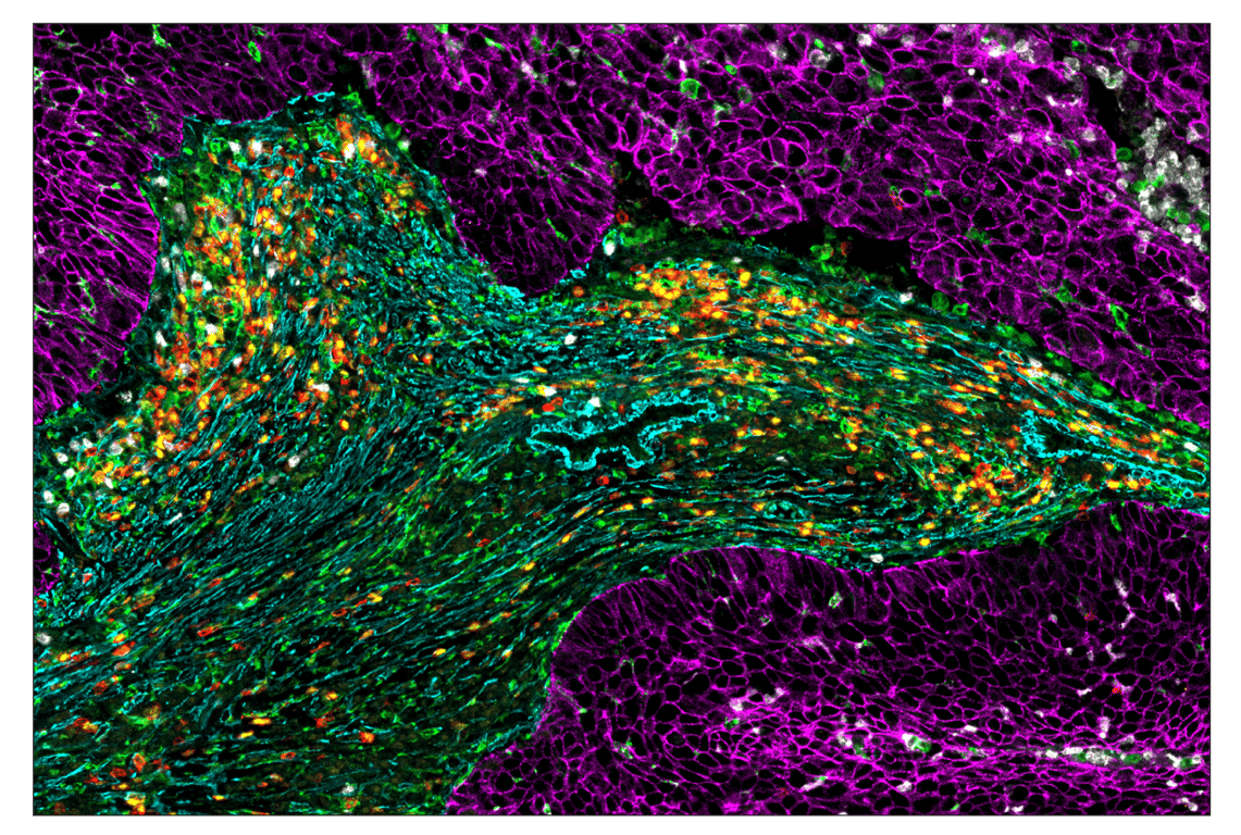
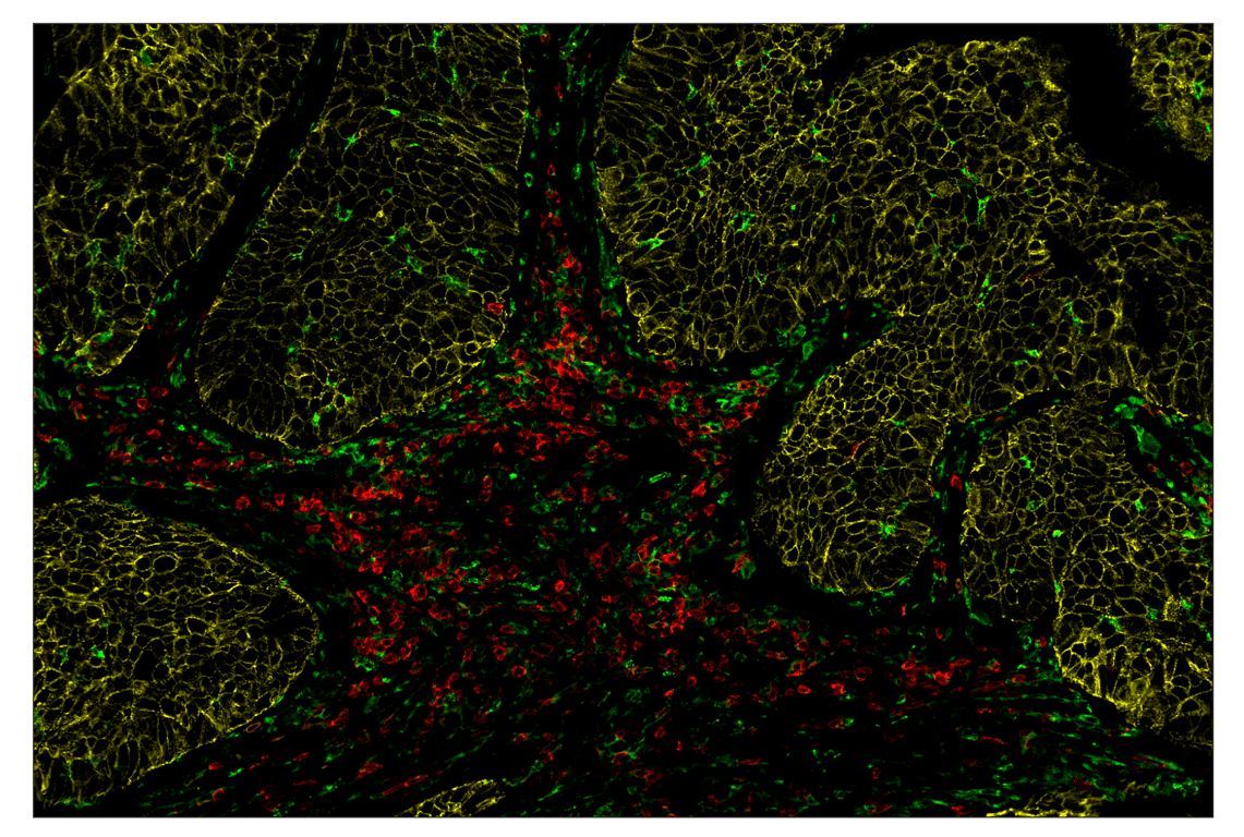
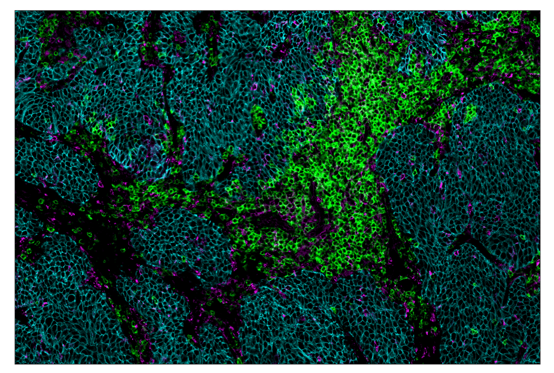
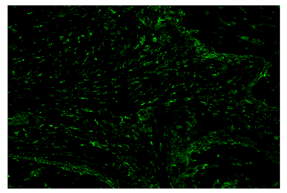
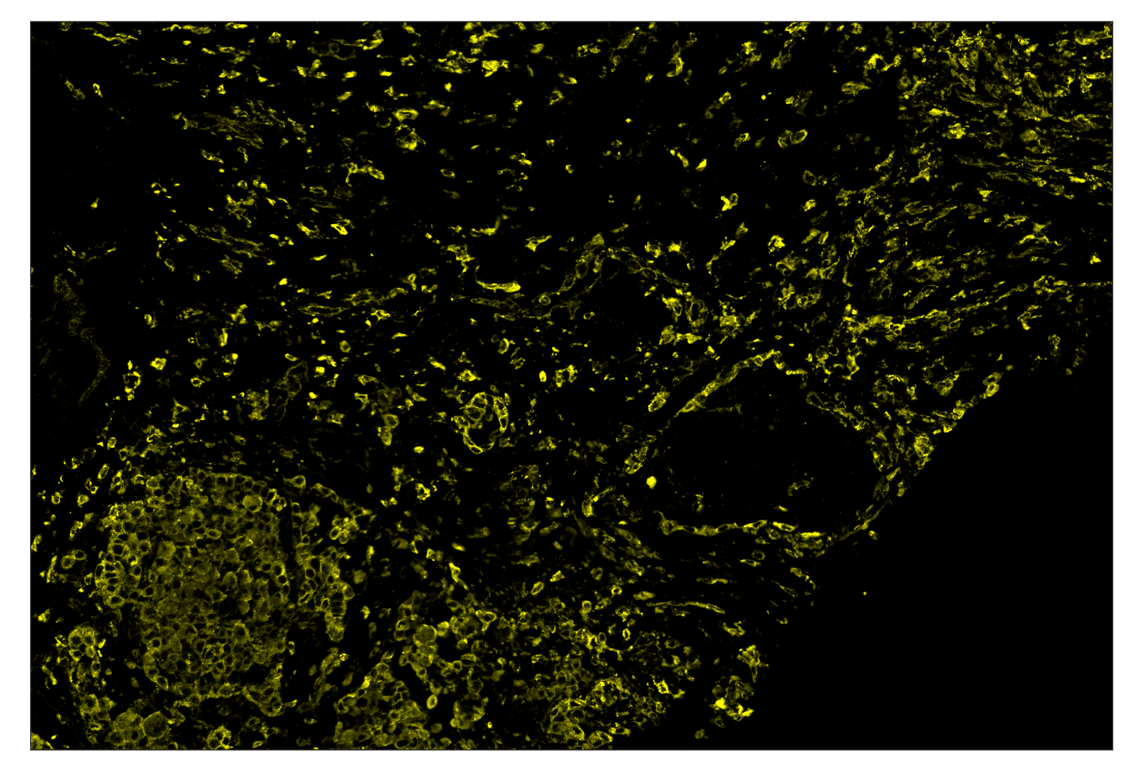
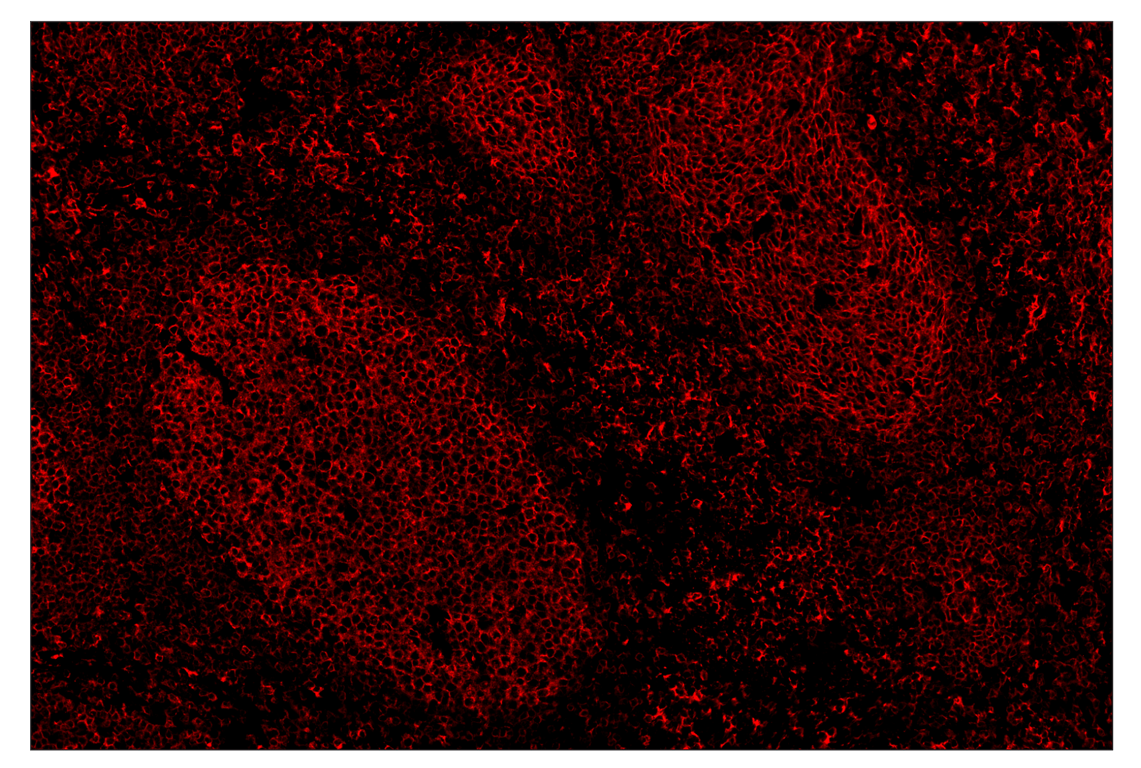
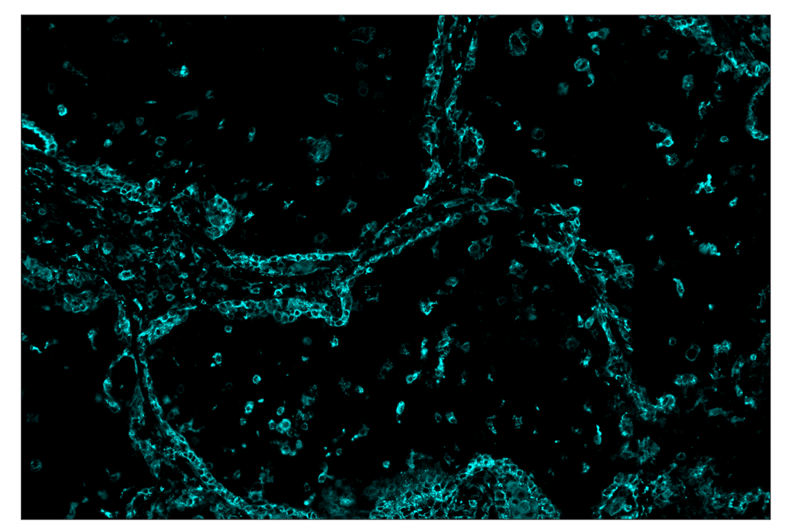
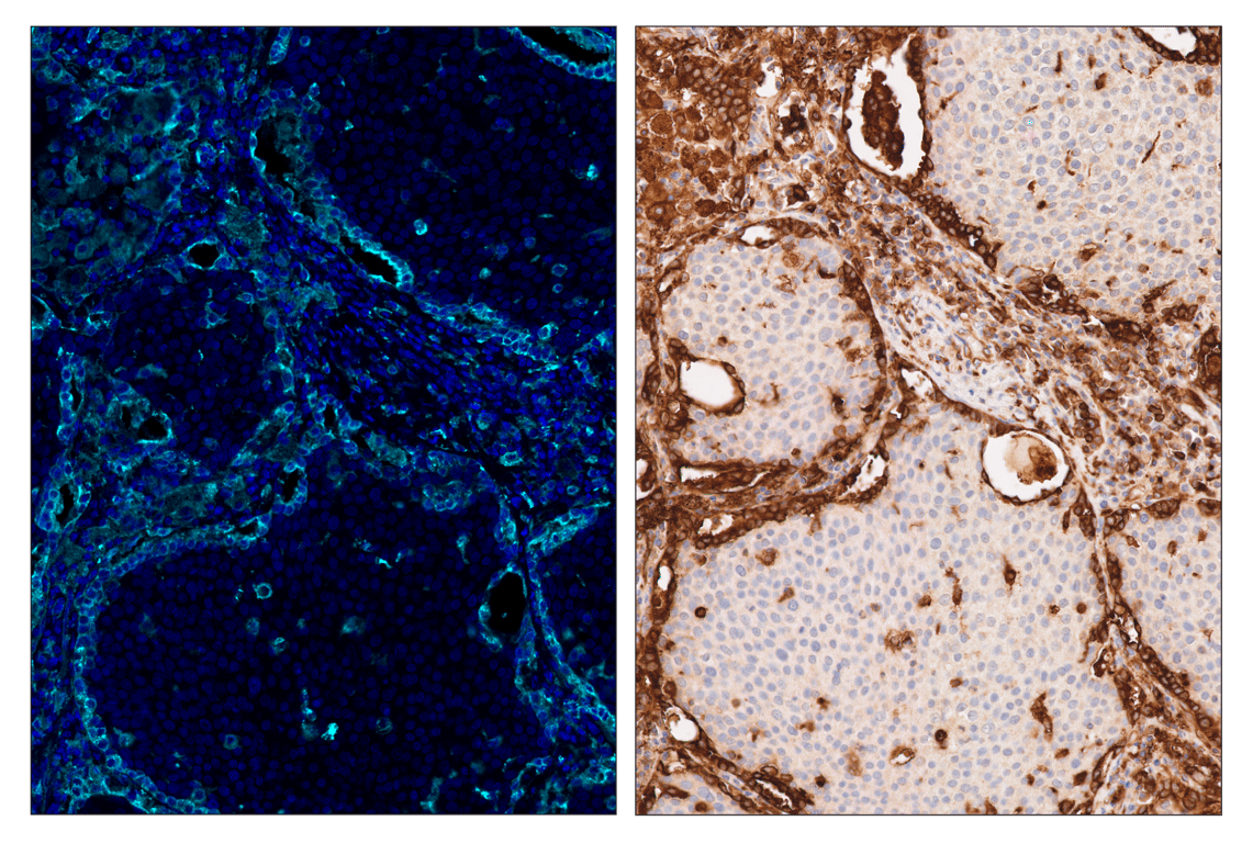
MHC Class II (LGII-612.14) & CO-0105-594 SignalStar™ Oligo-Antibody Pair #90033 (yellow). All fluorophores have been assigned a pseudocolor, as indicated. Staining was performed on the BOND RX autostainer by Leica Biosystems.
Immunohistochemistry Image 3: MHC Class II (LGII-612.14) & CO-0105-594 SignalStar<sup>™</sup> Oligo-Antibody Pair
Orders: 877-616-CELL (2355) orders@cellsignal.com Support: 877-678-TECH (8324) info@cellsignal.com Web: cellsignal.com
For Research Use Only. Not for Use in Diagnostic Procedures.

Revision 1

Cell Signaling Technology Logo
SignalStar™ multiplex immunohistochemical analysis of paraffin-embedded human tonsil using MHC Class II (LGII-612.14) & CO-0105-647 SignalStar™ Oligo-Antibody Pair #73441 (red). All fluorophores have been assigned a pseudocolor, as indicated. Staining was performed on the BOND RX autostainer by Leica Biosystems.
Immunohistochemistry Image 4: MHC Class II (LGII-612.14) & CO-0105-594 SignalStar<sup>™</sup> Oligo-Antibody Pair
SignalStar™ multiplex immunohistochemical analysis of paraffin-embedded human squamous cell lung carcinoma using MHC Class II (LGII-612.14) & CO-0105-750 SignalStar™ Oligo-Antibody Pair #23923 (cyan). All fluorophores have been assigned a pseudocolor, as indicated. Staining was performed on the BOND RX autostainer by Leica Biosystems.
Immunohistochemistry Image 5: MHC Class II (LGII-612.14) & CO-0105-594 SignalStar<sup>™</sup> Oligo-Antibody Pair
SignalStar™ multiplex immunohistochemical analysis of paraffin-embedded human squamous cell lung carcinoma using MHC Class II (LGII-612.14) & CO-0105-750 SignalStar™ Oligo-Antibody Pair #23923 (left, cyan) and DAPI #4083 (left, blue) compared to chromogenic immunohistochemical analysis of a serial section of paraffin-embedded human squamous cell lung carcinoma using MHC Class II (LGII-612.14) Mouse mAb #68258 (right). All fluorophores have been assigned a pseudocolor, as indicated. Staining was performed on the BOND RX autostainer by Leica Biosystems.
Immunohistochemistry Image 6: MHC Class II (LGII-612.14) & CO-0105-594 SignalStar<sup>™</sup> Oligo-Antibody Pair
Orders: 877-616-CELL (2355) orders@cellsignal.com Support: 877-678-TECH (8324) info@cellsignal.com Web: cellsignal.com
For Research Use Only. Not for Use in Diagnostic Procedures.

Revision 1

Cell Signaling Technology Logo
SignalStar™ multiplex immunohistochemical analysis of paraffin-embedded human squamous cell lung carcinoma using MHC Class II (LGII-612.14) & CO-0105-488 SignalStar™ Oligo-Antibody Pair #55436 (green), E-Cadherin (24E10) & CO-0103-594 SignalStar™ Oligo-Antibody Pair #65400 (magenta), CD28 (D2Z4E) & CO-0081-647 SignalStar™ Oligo-Antibody Pair #96977 (red), α-Smooth Muscle Actin (D4K9N) & CO-0024-488 SignalStar™ Oligo-Antibody Pair #76133 (green), TCF1/TCF7 (C63D9) & CO-0006-594 SignalStar™ Oligo-Antibody Pair #34706 (yellow), and Arginase-1 (D4E3M) & CO-0075-647 SignalStar™ Oligo-Antibody Pair #44900 (white). All fluorophores have been assigned a pseudocolor, as indicated. Staining was performed on the BOND RX autostainer by Leica Biosystems.
Immunohistochemistry Image 7: MHC Class II (LGII-612.14) & CO-0105-594 SignalStar<sup>™</sup> Oligo-Antibody Pair
SignalStar™ multiplex immunohistochemical analysis of paraffin-embedded human squamous cell lung carcinoma using MHC Class II (LGII-612.14) & CO-0105-488 SignalStar™ Oligo-Antibody Pair #55436 (green), E-Cadherin (24E10) & CO-0103-594 SignalStar™ Oligo-Antibody Pair #65400 (yellow), and CD28 (D2Z4E) & CO-0081-647 SignalStar™ Oligo-Antibody Pair #96977 (red). All fluorophores have been assigned a pseudocolor, as indicated. 
Immunohistochemistry Image 1: MHC Class II (LGII-612.14) & CO-0105-594 SignalStar<sup>™</sup> Oligo-Antibody Pair
Orders: 877-616-CELL (2355) orders@cellsignal.com Support: 877-678-TECH (8324) info@cellsignal.com Web: cellsignal.com
For Research Use Only. Not for Use in Diagnostic Procedures.