Revision 4
#9653
Store at -20C
Double Strand Breaks (DSB) Repair Antibody Sampler Kit
1 Kit
(9 x 20 microliters)
877-616-CELL (2355)
877-678-TECH (8324)
3 Trask Lane | Danvers | Massachusetts | 01923 | USA
For Research Use Only. Not for Use in Diagnostic Procedures.
| Product Includes | Product # | Quantity | Mol. Wt | Isotype/Source |
|---|---|---|---|---|
| Phospho-ATM (Ser1981) (D25E5) Rabbit mAb | 13050 | 20 µl | 350 kDa | Rabbit IgG |
| ATM (D2E2) Rabbit mAb | 2873 | 20 µl | 350 kDa | Rabbit IgG |
| Phospho-BRCA1 (Ser1524) Antibody | 9009 | 20 µl | 220 kDa | Rabbit |
| DNA-PKcs Antibody | 4602 | 20 µl | 450 kDa | Rabbit |
| Ku80 (C48E7) Rabbit mAb | 2180 | 20 µl | 86 kDa | Rabbit IgG |
| Mre11 (31H4) Rabbit mAb | 4847 | 20 µl | 81 kDa | Rabbit IgG |
| Phospho-p95/NBS1 (Ser343) Antibody | 3001 | 20 µl | 95 kDa | Rabbit |
| Rad50 Antibody | 3427 | 20 µl | 153 kDa | Rabbit |
| XLF Antibody | 2854 | 20 µl | 39 kDa | Rabbit |
| Anti-rabbit IgG, HRP-linked Antibody | 7074 | 100 µl | Goat |
Please visit cellsignal.com for individual component applications, species cross-reactivity, dilutions, protocols, and additional product information.
Description
The Double Strand Breaks (DSB) Repair Antibody Sampler Kit provides an economical means to investigate repair of double-strand DNA breaks within the cell. The kit contains primary and secondary antibodies to perform two western blots with each antibody.
Storage
Background
Double strand DNA breaks (DSB) in mammalian cells can be repaired by the related mechanisms of non-homologous end-joining (NHEJ) and homologous recombination (HR). A DNA-dependent protein kinase composed of DNA-binding subunits Ku70 and Ku86 and the DNA-PKcs catalytic subunit mediates NHEJ repair. The Ku heterodimer binds free DNA ends and recruits DNA-PKcs to the break (1). DNA-PKcs signals areas of DNA damage and recruits additional proteins, such as the Artemis exo- and endonuclease that processes and primes the damaged sequence (2,3). Following replacement DNA synthesis, a ligase complex composed of DNA ligase IV and XRCC4 joins the repaired ends. XRCC4-like factor (XLF) is an essential ligase-associated repair factor that promotes gap-filling during NHEJ (4). Homologous recombination utilizes aligned homologous sequences as a repair template. The MRN complex, composed of Mre11, Rad50, and nibrin (p95/NBS1), plays a critical role in sensing, processing and repairing breaks (5). MRN interacts with BRCA1 and CtIP to facilitate 5’ resection of DSB DNA to generate 3’ ssDNA ends necessary for repair (6). DNA-binding protein Mre11 exhibits exonuclease and endonuclease activity and is largely responsible for ssDNA end processing (7). Interaction between the MRN complex and ATM kinase promotes association between the kinase and its substrates and likely leads to ATM activation (8). ATM acts a central controller of the cell cycle checkpoint by phosphorylating multiple targets, including c-Abl, BRCA1 and p95/NSB1. Activated c-Abl phosphorylates Rad52, which promotes Rad51 binding to ssDNA and subsequent annealing of ssDNA (7).
Background References
- Gottlieb, T.M. and Jackson, S.P. (1993) Cell 72, 131-42.
- Franco, S. et al. (2008) J Exp Med 205, 557-64.
- Collis, S.J. et al. (2005) Oncogene 24, 949-61.
- Akopiants, K. et al. (2009) Nucleic Acids Res 37, 4055-62.
- Williams, R.S. et al. (2007) Biochem Cell Biol 85, 509-20.
- Chen, L. et al. (2008) J Biol Chem 283, 7713-20.
- Czornak, K. et al. (2008) J Appl Genet 49, 383-96.
- Lee, J.H. and Paull, T.T. (2007) Oncogene 26, 7741-8.
Trademarks and Patents
Cell Signaling Technology is a trademark of Cell Signaling Technology, Inc.
All other trademarks are the property of their respective owners. Visit cellsignal.com/trademarks for more information.
Limited Uses
Except as otherwise expressly agreed in a writing signed by a legally authorized representative of CST, the following terms apply to Products provided by CST, its affiliates or its distributors. Any Customer's terms and conditions that are in addition to, or different from, those contained herein, unless separately accepted in writing by a legally authorized representative of CST, are rejected and are of no force or effect.
Products are labeled with For Research Use Only or a similar labeling statement and have not been approved, cleared, or licensed by the FDA or other regulatory foreign or domestic entity, for any purpose. Customer shall not use any Product for any diagnostic or therapeutic purpose, or otherwise in any manner that conflicts with its labeling statement. Products sold or licensed by CST are provided for Customer as the end-user and solely for research and development uses. Any use of Product for diagnostic, prophylactic or therapeutic purposes, or any purchase of Product for resale (alone or as a component) or other commercial purpose, requires a separate license from CST. Customer shall (a) not sell, license, loan, donate or otherwise transfer or make available any Product to any third party, whether alone or in combination with other materials, or use the Products to manufacture any commercial products, (b) not copy, modify, reverse engineer, decompile, disassemble or otherwise attempt to discover the underlying structure or technology of the Products, or use the Products for the purpose of developing any products or services that would compete with CST products or services, (c) not alter or remove from the Products any trademarks, trade names, logos, patent or copyright notices or markings, (d) use the Products solely in accordance with CST Product Terms of Sale and any applicable documentation, and (e) comply with any license, terms of service or similar agreement with respect to any third party products or services used by Customer in connection with the Products.
Revision 4
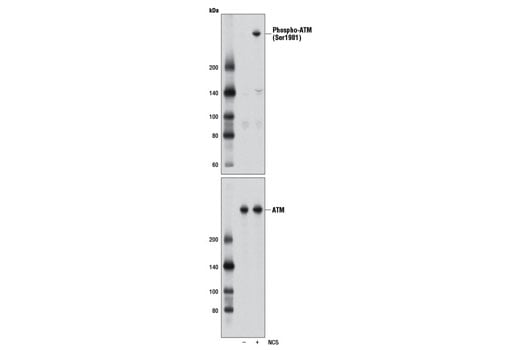
Western blot analysis of extracts from HCT 116 cells, untreated (-) or treated with neocarzinostatin (NCS 10 μM, 1 hr; +), using Phospho-ATM (Ser1981) (D25E5) Rabbit mAb (upper) and ATM (D2E2) Rabbit mAb #2873 (lower).
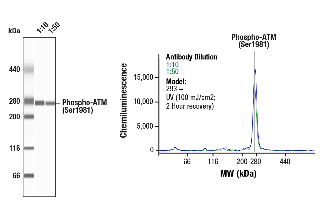
Simple Western™ analysis of lysates (0.1 mg/mL) from 293 cells treated with UV (100 mJ/cm2; 2 Hour Recovery) using Phospho-ATM (Ser1981) (D25E5) Rabbit mAb #13050. The virtual lane view (left) shows the target band (as indicated) at 1:10 and 1:50 dilutions of primary antibody. The corresponding electropherogram view (right) plots chemiluminescence by molecular weight along the capillary at 1:10 (blue line) and 1:50 (green line) dilutions of primary antibody. This experiment was performed under reducing conditions on the Jess™ Simple Western instrument from ProteinSimple, a BioTechne brand, using the 66 - 440 kDa separation module.
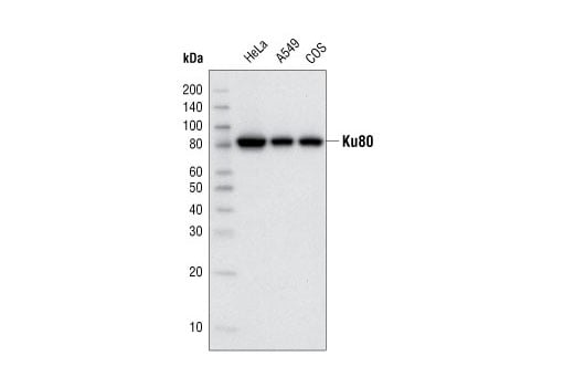
Western blot analysis of cell extracts from HeLa, A549 and COS cells using Ku80 (C48E7) Rabbit mAb.
Revision 4
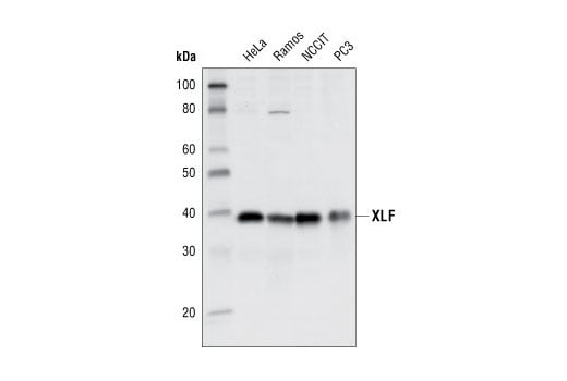
Western blot analysis of extracts of various cell lines using XLF Antibody.
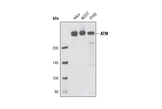
Western blot analysis of extracts of HeLa, NCCIT and PYS2 cells using ATM (D2E2) Rabbit mAb.
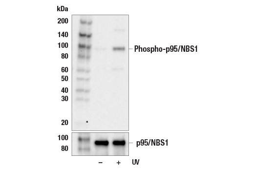
Western blot analysis of extracts from 293 cells, untreated or UV-treated (100 mJ/cm2, 1 hr), using Phospho-p95/NBS1 (Ser343) Antibody (upper) or p95/NBS1 (E8M3Q) XP® Rabbit mAb #81234 (lower).
Revision 4
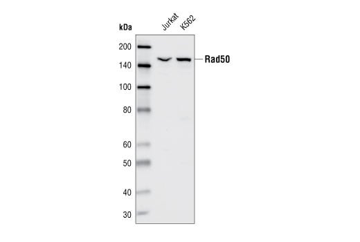
Western blot analysis of extracts from Jurkat and K562 cells, using RAD50 Antibody.
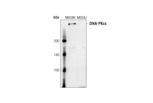
Western blot analysis of extracts from M059K (DNA-PK wildtype) and M059J (DNA-PK deficient) cells, using DNA-PK Antibody.
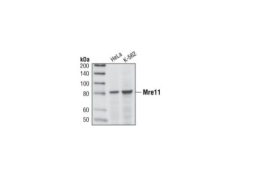
Western blot analysis of extracts from HeLa and K562 cells, using Mre11 Rabbit (31H4) mAb.
Revision 4
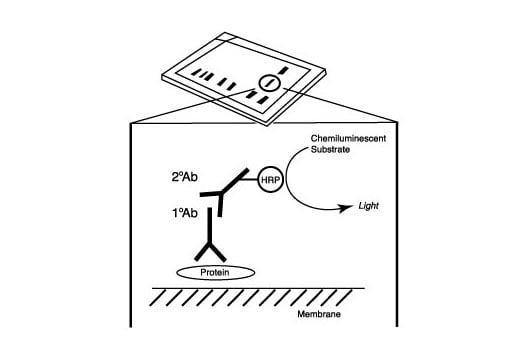
After the primary antibody is bound to the target protein, a complex with HRP-linked secondary antibody is formed. The LumiGLO® is added and emits light during enzyme catalyzed decomposition.
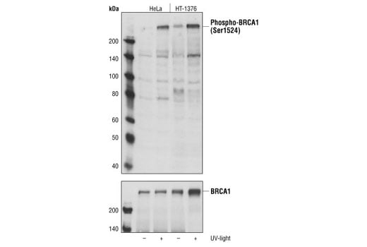
Western blot analysis of untreated and UV-treated (50 mJ/cm2,30 min) HeLa cells and HT-1376 cells, using Phospho-BRCA1 (Ser1524) Antibody (upper) and BRCA1 Antibody #9010 (lower).
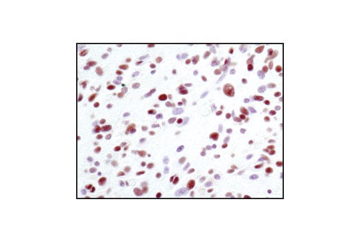
Immunohistochemical analysis of paraffin-embedded human glioblastoma using Ku80 (C48E7) Rabbit mAb.
Revision 4
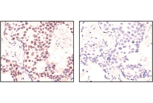
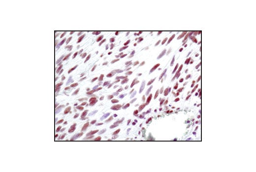
Immunohistochemical analysis of paraffin-embedded human breast carcinoma, using Mre11 (31H4) Rabbit mAb in the presence of control peptide (left) or Mre11 Blocking Peptide #1035 (right).
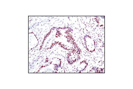
Immunohistochemical analysis of paraffin-embedded human colon carcinoma using Ku80 (C48E7) Rabbit mAb.
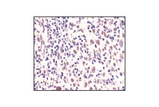
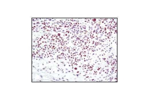
Immunohistochemical analysis of paraffin-embedded lung carcinoma, using Mre11 (31H4) Rabbit mAb.
Revision 4
Immunohistochemical analysis of paraffin-embedded human melanoma using Ku80 (C48E7) Rabbit mAb.
Immunohistochemical analysis of paraffin-embedded human GIST using Ku80 (C48E7) Rabbit mAb.
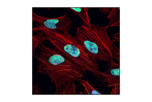
Confocal immunofluorescent analysis of HeLa cells using Ku80 (C48E7) Rabbit mAb (green). Actin filaments have been labeled with Alexa Fluor® 555 phalloidin (red).
Revision 4
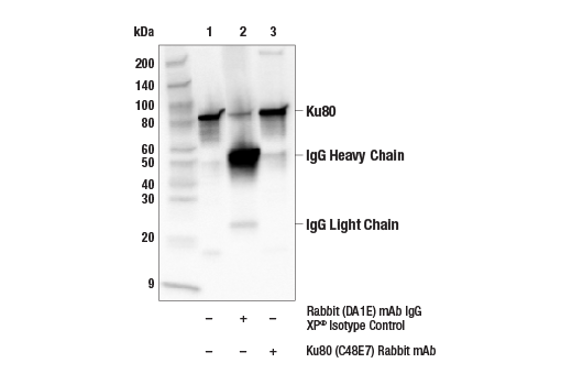
Immunoprecipitation of Ku80 from HeLa cells. Lane 1 is 10% input, lane 2 is precipitated with Rabbit (DA1E) mAb IgG XP® Isotype Control #3900, and lane 3 is Ku80 (C48E7) Rabbit mAb, #2180. Western blot was performed using Ku80 (C48E7) Rabbit mAb, #2180.