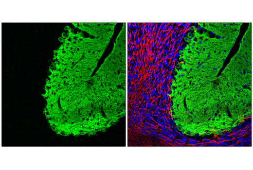
WB, IF-F
H M R
Endogenous
20, 22, 50, 70, 80
Rabbit IgG
#P48664
6511
Product Information
Product Usage Information
| Application | Dilution |
|---|---|
| Western Blotting | 1:1000 |
| Immunofluorescence (Frozen) | 1:200 - 1:400 |
Storage
Specificity / Sensitivity
Species Reactivity:
Human, Mouse, Rat
Source / Purification
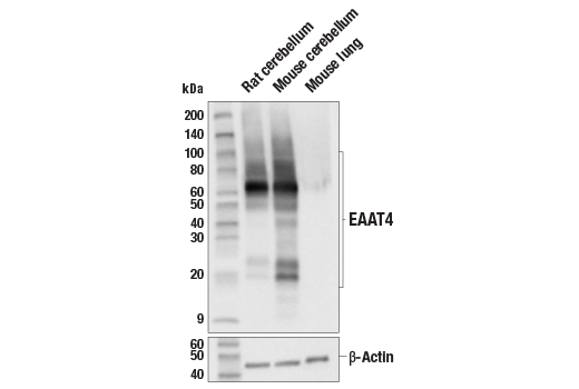
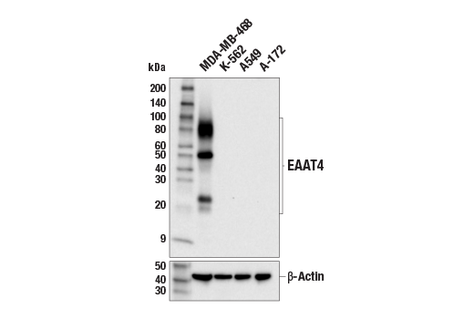
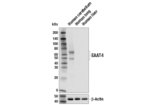
Monoclonal antibody is produced by immunizing animals with a synthetic peptide corresponding to residues surrounding Gly552 of human EAAT4 protein.
Background
During neurotransmission, glutamate is released from vesicles of the presynaptic cell, and glutamate receptors (e.g., NMDA receptor, AMPA receptor) bind glutamate for activation at the opposing postsynaptic cell. Excitatory amino acid transporters (EAATs) regulate and maintain extracellular glutamate concentrations below excitotoxic levels (1,2). In addition, glutamate transporters may limit the duration of synaptic excitation by an electrogenic process in which the transmitter is cotransported with three sodium ions and one proton, followed by countertransport of a potassium ion (1,2). Five EAATs (EAAT1-5) have been identified. EAAT1 and EAAT2 are expressed mainly in glia, while EAAT3, EAAT4, and EAAT5 are considered to be neuronal transporters (2). EAAT4 (encoded by the SLC1A6 gene) is a neuronal glutamate transporter expressed in Purkinje cells of the cerebellum (3). EAAT4 plays an important role in maintaining neuronal synaptic function, particularly in the cerebellum, by maintaining appropriate extracellular glutamate levels to prevent neurotoxicity and permit effective synaptic communication. EAAT4 function may be regulated by post-translational modification (4,5). EAAT4 may be a therapeutic target to treat several neurological diseases, including spinocerebellar ataxias, by restoring extracellular glutamate homeostasis (6).
- Amara, S.G. and Fontana, A.C. (2002) Neurochem Int 41, 313-8.
- Tanaka, K. et al. (1997) Science 276, 1699-702.
- Yamada, K. et al. (1996) Neuroreport 7, 2013-7.
- Böhmer, C. et al. (2004) Biochem Biophys Res Commun 324, 1242-8.
- Alesutan, I.S. et al. (2010) Cell Physiol Biochem 25, 187-94.
- Perkins, E.M. et al. (2018) Hum Mol Genet 27, 2614-2627.
Species Reactivity
Species reactivity is determined by testing in at least one approved application (e.g., western blot).
Western Blot Buffer
IMPORTANT: For western blots, incubate membrane with diluted primary antibody in 5% w/v BSA, 1X TBS, 0.1% Tween® 20 at 4°C with gentle shaking, overnight.
Applications Key
WB: Western Blotting IF-F: Immunofluorescence (Frozen)
Cross-Reactivity Key
H: human M: mouse R: rat Hm: hamster Mk: monkey Vir: virus Mi: mink C: chicken Dm: D. melanogaster X: Xenopus Z: zebrafish B: bovine Dg: dog Pg: pig Sc: S. cerevisiae Ce: C. elegans Hr: horse GP: Guinea Pig Rab: rabbit All: all species expected
Trademarks and Patents
Limited Uses
Except as otherwise expressly agreed in a writing signed by a legally authorized representative of CST, the following terms apply to Products provided by CST, its affiliates or its distributors. Any Customer's terms and conditions that are in addition to, or different from, those contained herein, unless separately accepted in writing by a legally authorized representative of CST, are rejected and are of no force or effect.
Products are labeled with For Research Use Only or a similar labeling statement and have not been approved, cleared, or licensed by the FDA or other regulatory foreign or domestic entity, for any purpose. Customer shall not use any Product for any diagnostic or therapeutic purpose, or otherwise in any manner that conflicts with its labeling statement. Products sold or licensed by CST are provided for Customer as the end-user and solely for research and development uses. Any use of Product for diagnostic, prophylactic or therapeutic purposes, or any purchase of Product for resale (alone or as a component) or other commercial purpose, requires a separate license from CST. Customer shall (a) not sell, license, loan, donate or otherwise transfer or make available any Product to any third party, whether alone or in combination with other materials, or use the Products to manufacture any commercial products, (b) not copy, modify, reverse engineer, decompile, disassemble or otherwise attempt to discover the underlying structure or technology of the Products, or use the Products for the purpose of developing any products or services that would compete with CST products or services, (c) not alter or remove from the Products any trademarks, trade names, logos, patent or copyright notices or markings, (d) use the Products solely in accordance with CST Product Terms of Sale and any applicable documentation, and (e) comply with any license, terms of service or similar agreement with respect to any third party products or services used by Customer in connection with the Products.