Revision 2
#37283
Store at +4C
877-616-CELL (2355)
877-678-TECH (8324)
3 Trask Lane | Danvers | Massachusetts | 01923 | USA
For Research Use Only. Not for Use in Diagnostic Procedures.
Applications:
W, IHC-Bond, IHC-P
Reactivity:
H
Sensitivity:
Endogenous
MW (kDa):
38
Source/Isotype:
Mouse IgG1
UniProt ID:
#P15328
Entrez-Gene Id:
2348
Product Usage Information
| Application | Dilution |
|---|---|
| Western Blotting | 1:1000 |
| IHC Leica Bond | 1:50 - 1:200 |
| Immunohistochemistry (Paraffin) | 1:50 - 1:200 |
Storage
Specificity/Sensitivity
Folate Receptor Alpha/FOLR1 (BN3.2) Mouse mAb recognizes endogenous levels of FOLR1 protein.
Source / Purification
Monoclonal antibody is produced by immunizing animals with a prokaryotic recombinant protein corresponding to 189 amino acids of the external domain of human FOLR1 protein.
Background
Folate Receptor Alpha, also called FOLR1, is a cysteine-rich cell surface receptor that plays a critical role in the maintenance and transport of folate (vitamin B9) and reduced folic acid derivatives into cells and is essential for various cellular processes (1,2). FOLR1 binds to folate and facilitates its internalization into cells through receptor-mediated endocytosis or potocytosis and is important in cellular processes, including DNA synthesis and repair (1-3). Intracellular folate levels are critical to maintain and support folate functions in its multiple coenzyme forms (4). It is primarily expressed in tissues requiring high folate levels, such as the placenta, kidneys, and certain cancer cells (5). FOLR1 has been found to be overexpressed in several types of cancer, including ovarian, lung, and breast cancer (6-8).
Background References
- Chen, C. et al. (2013) Nature 500, 486-9.
- Wibowo, A.S. et al. (2013) Proc Natl Acad Sci USA 110, 15180-8.
- Orr, R.B. and Kamen, B.A. (1994) Cancer Res 54, 3905-11.
- Bailey, L.B. and Gregory, J.F. (1999) J Nutr 129, 779-82.
- Lacey, S.W. et al. (1989) J Clin Invest 84, 715-20.
- Bax, H.J. et al. (2023) Br J Cancer 128, 342-353.
- Nunez, M.I. et al. (2012) J Thorac Oncol 7, 833-40.
- Necela, B.M. et al. (2015) PLoS One 10, e0122209.
Species Reactivity
Species reactivity is determined by testing in at least one approved application (e.g., western blot).
Western Blot Buffer
IMPORTANT: For western blots, incubate membrane with diluted primary antibody in 5% w/v BSA, 1X TBS, 0.1% Tween® 20 at 4°C with gentle shaking, overnight.
Applications Key
W: Western Blotting IHC-Bond: IHC Leica Bond
Cross-Reactivity Key
H: Human M: Mouse R: Rat Hm: Hamster Mk: Monkey Vir: Virus Mi: Mink C: Chicken Dm: D. melanogaster X: Xenopus Z: Zebrafish B: Bovine Dg: Dog Pg: Pig Sc: S. cerevisiae Ce: C. elegans Hr: Horse GP: Guinea Pig Rab: Rabbit G: Goat All: All Species Expected
Trademarks and Patents
Cell Signaling Technology is a trademark of Cell Signaling Technology, Inc.
All other trademarks are the property of their respective owners. Visit cellsignal.com/trademarks for more information.
Limited Uses
Except as otherwise expressly agreed in a writing signed by a legally authorized representative of CST, the following terms apply to Products provided by CST, its affiliates or its distributors. Any Customer's terms and conditions that are in addition to, or different from, those contained herein, unless separately accepted in writing by a legally authorized representative of CST, are rejected and are of no force or effect.
Products are labeled with For Research Use Only or a similar labeling statement and have not been approved, cleared, or licensed by the FDA or other regulatory foreign or domestic entity, for any purpose. Customer shall not use any Product for any diagnostic or therapeutic purpose, or otherwise in any manner that conflicts with its labeling statement. Products sold or licensed by CST are provided for Customer as the end-user and solely for research and development uses. Any use of Product for diagnostic, prophylactic or therapeutic purposes, or any purchase of Product for resale (alone or as a component) or other commercial purpose, requires a separate license from CST. Customer shall (a) not sell, license, loan, donate or otherwise transfer or make available any Product to any third party, whether alone or in combination with other materials, or use the Products to manufacture any commercial products, (b) not copy, modify, reverse engineer, decompile, disassemble or otherwise attempt to discover the underlying structure or technology of the Products, or use the Products for the purpose of developing any products or services that would compete with CST products or services, (c) not alter or remove from the Products any trademarks, trade names, logos, patent or copyright notices or markings, (d) use the Products solely in accordance with CST Product Terms of Sale and any applicable documentation, and (e) comply with any license, terms of service or similar agreement with respect to any third party products or services used by Customer in connection with the Products.
Revision 2
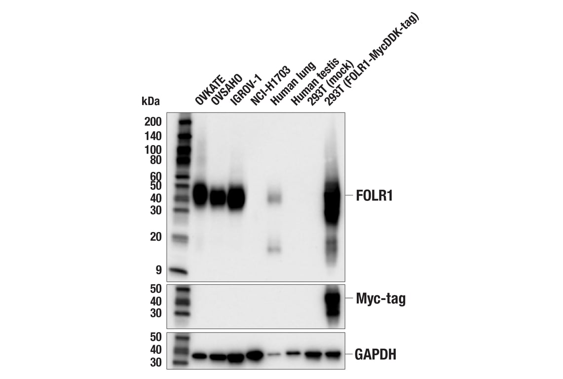
Western blot analysis of extracts from various cells and tissues using Folate Receptor Alpha/FOLR1 (BN3.2) Mouse mAb (upper), Myc-Tag (71D10) Rabbit mAb #2278 (middle), or GAPDH (D16H11) XP® Rabbit mAb #5174 (lower).
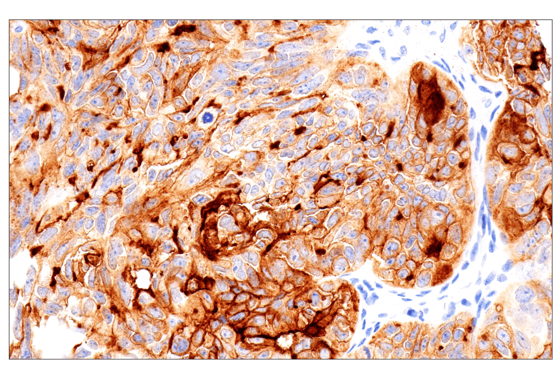
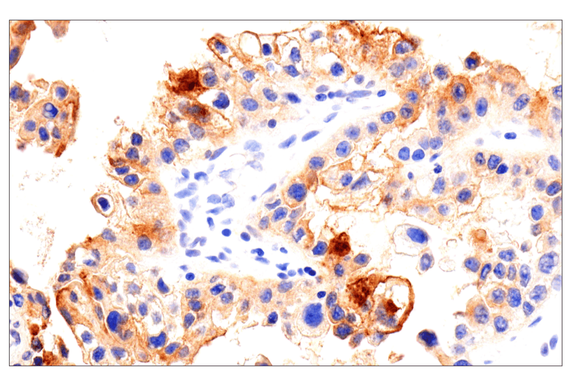
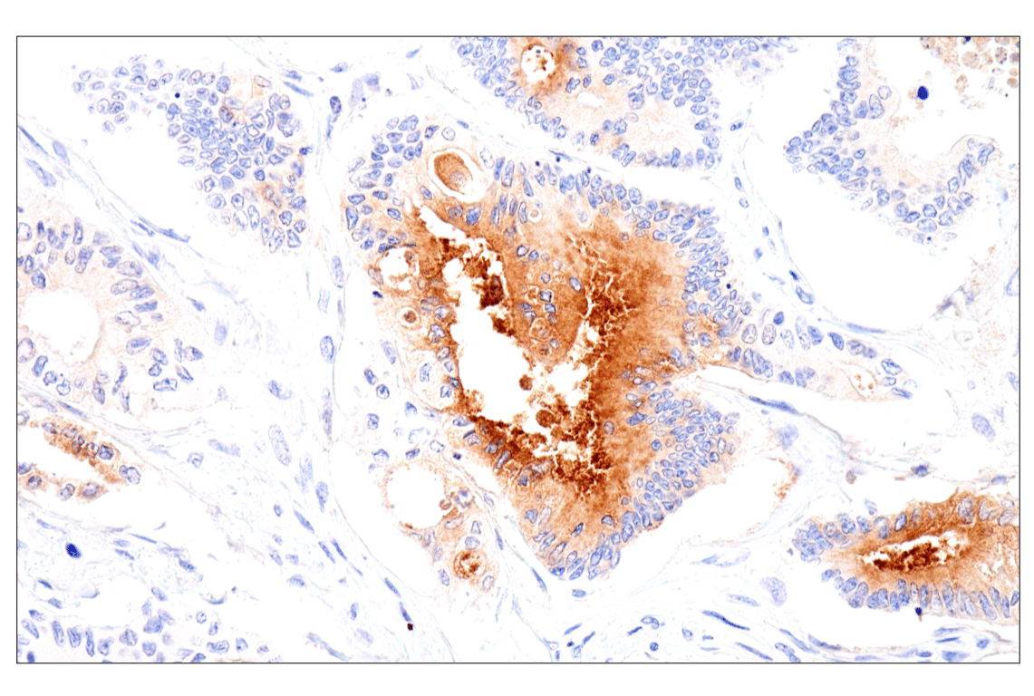
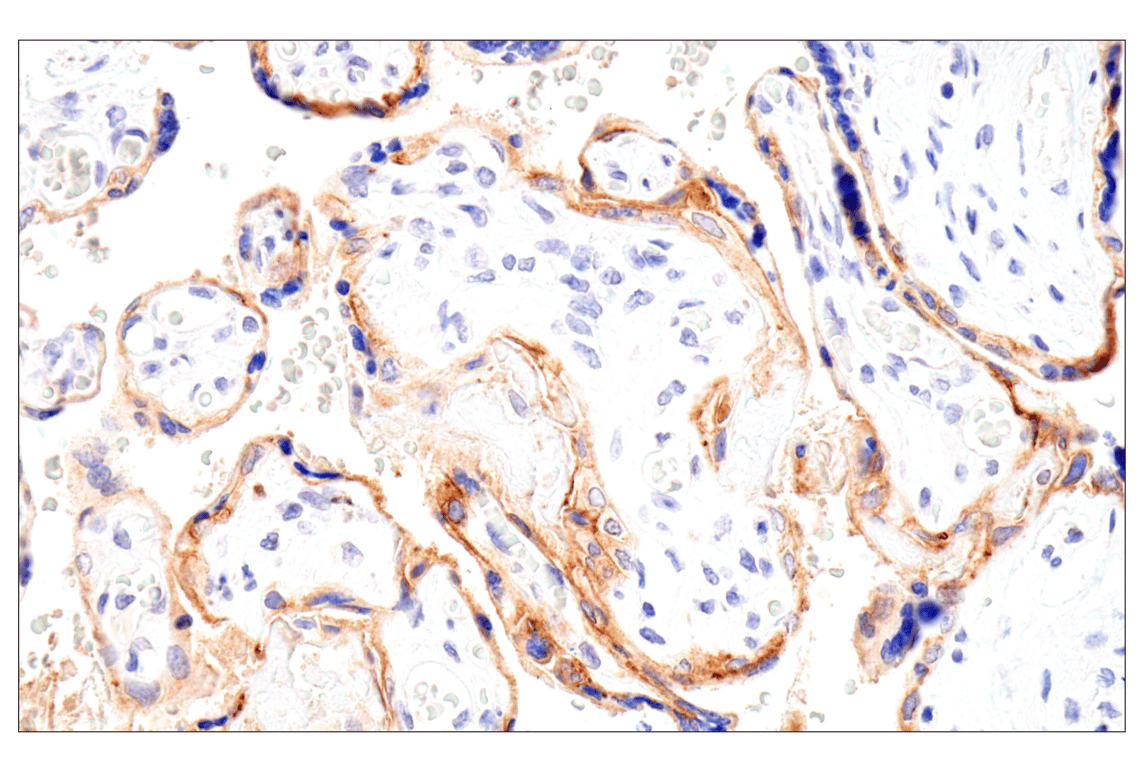
Immunohistochemical analysis of paraffin-embedded human serous papillary carcinoma of the ovary using Folate Receptor Alpha/FOLR1 (BN3.2) Mouse mAb performed on the Leica BOND RX.
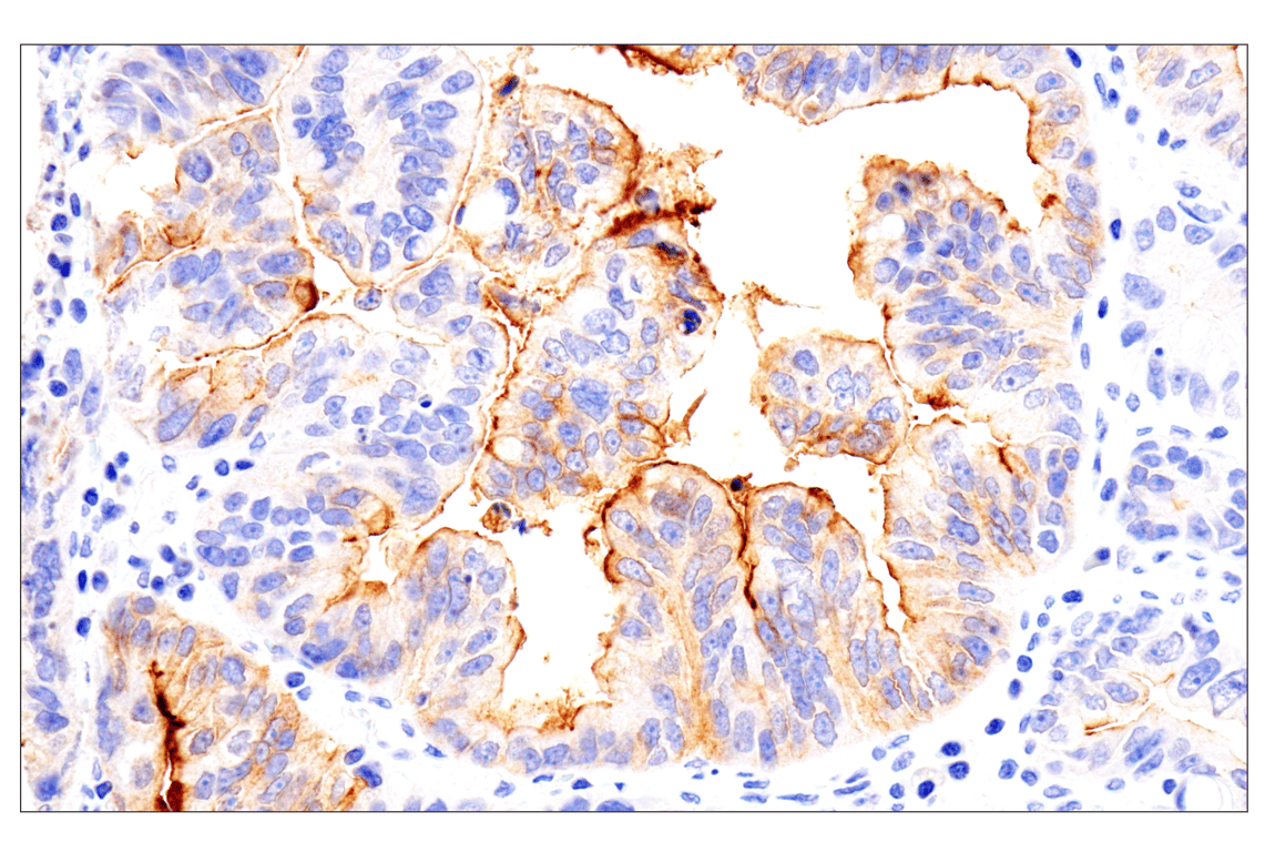
Immunohistochemical analysis of paraffin-embedded human esophageal adenocarcinoma using Folate Receptor Alpha/FOLR1 (BN3.2) Mouse mAb performed on the Leica BOND RX.
Revision 2
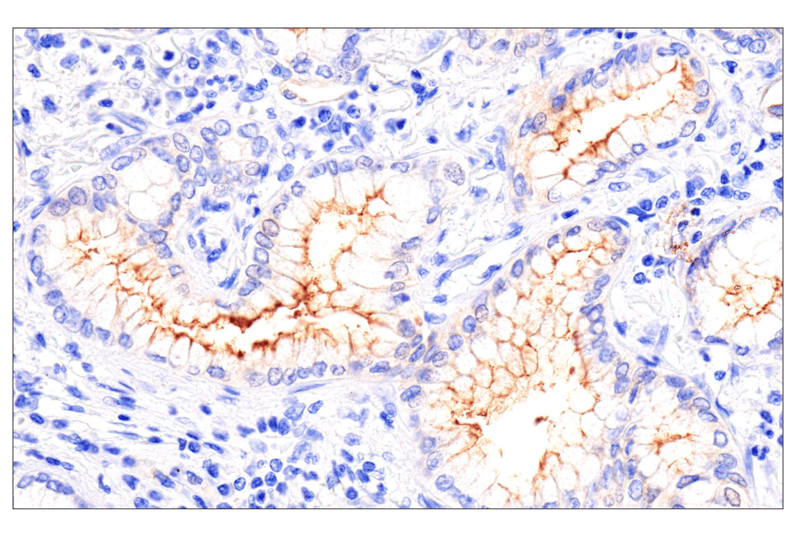
Immunohistochemical analysis of paraffin-embedded human bronchioloalveolar adenocarcinoma using Folate Receptor Alpha/FOLR1 (BN3.2) Mouse mAb performed on the Leica BOND RX.
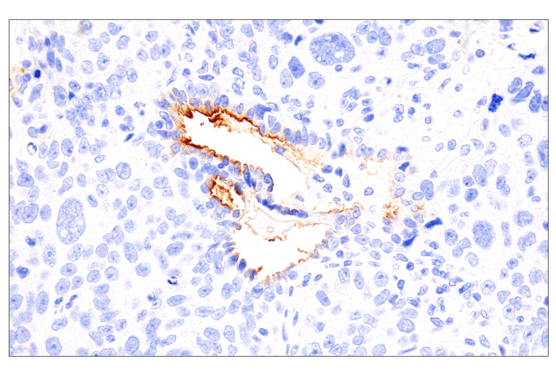
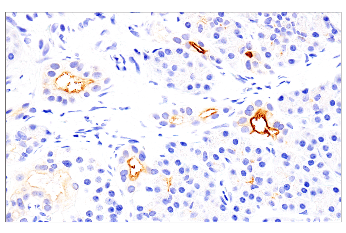
Immunohistochemical analysis of paraffin-embedded human renal cell carcinoma using Folate Receptor Alpha/FOLR1 (BN3.2) Mouse mAb performed on the Leica BOND RX.
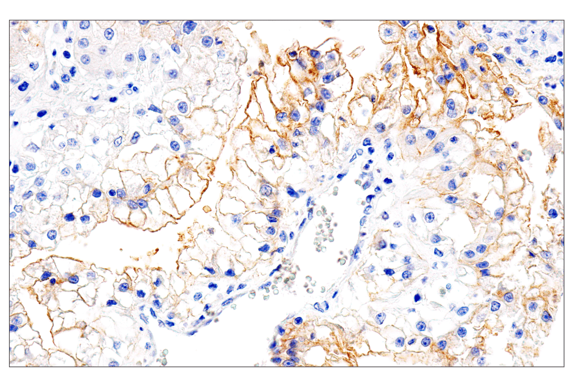
Immunohistochemical analysis of paraffin-embedded human ovarian clear cell carcinoma using Folate Receptor Alpha/FOLR1 (BN3.2) Mouse mAb performed on the Leica BOND RX.
Revision 2
Immunohistochemical analysis of paraffin-embedded human serous papillary carcinoma of the ovary using Folate Receptor Alpha/FOLR1 (BN3.2) Mouse mAb performed on the Leica BOND RX.
Immunohistochemical analysis of paraffin-embedded human squamous cell lung carcinoma using Folate Receptor Alpha/FOLR1 (BN3.2) Mouse mAb performed on the Leica BOND RX.
Immunohistochemical analysis of paraffin-embedded normal human pancreas using Folate Receptor Alpha/FOLR1 (BN3.2) Mouse mAb performed on the Leica BOND RX.
Revision 2
Immunohistochemical analysis of paraffin-embedded human pulmonary sarcoma using Folate Receptor Alpha/FOLR1 (BN3.2) Mouse mAb.
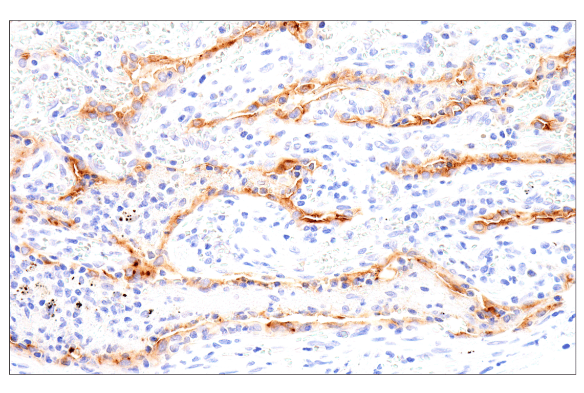
Immunohistochemical analysis of paraffin-embedded human colon adenocarcinoma using Folate Receptor Alpha/FOLR1 (BN3.2) Mouse mAb.
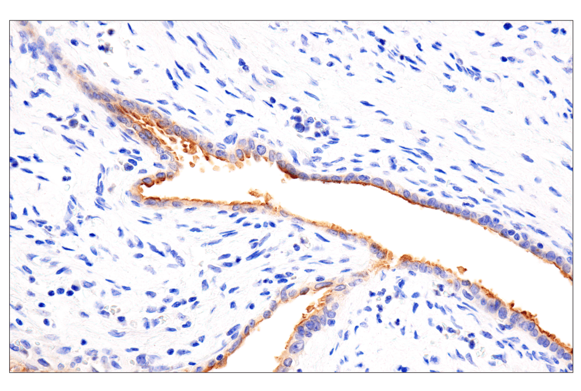
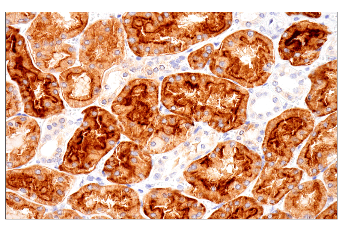
Immunohistochemical analysis of paraffin-embedded normal human kidney using Folate Receptor Alpha/FOLR1 (BN3.2) Mouse mAb.
Revision 2
Immunohistochemical analysis of paraffin-embedded normal human lung using Folate Receptor Alpha/FOLR1 (BN3.2) Mouse mAb.
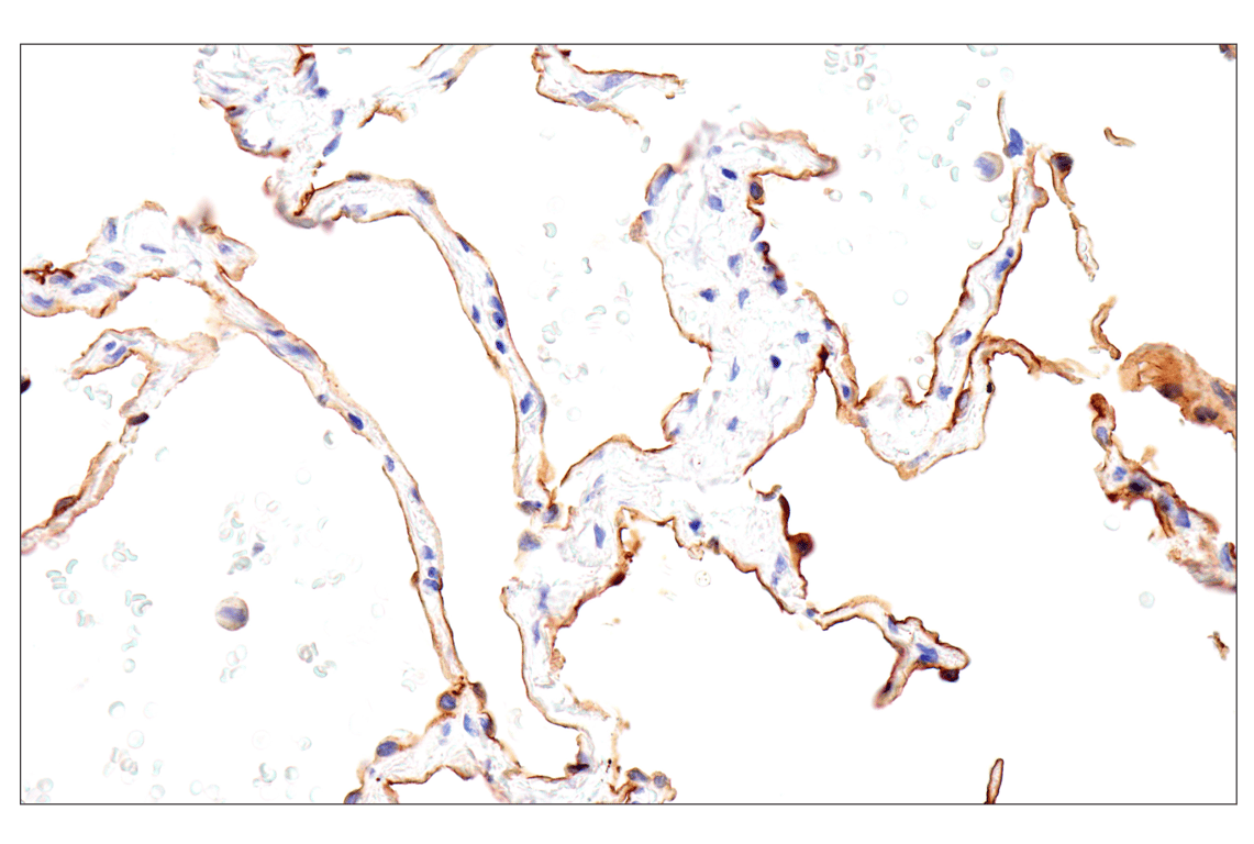
Immunohistochemical analysis of paraffin-embedded normal human placenta using Folate Receptor Alpha/FOLR1 (BN3.2) Mouse mAb.
Immunohistochemical analysis of paraffin-embedded human esophageal adenocarcinoma using Folate Receptor Alpha/FOLR1 (BN3.2) Mouse mAb (left) compared to concentration-matched Rabbit (DA1E) mAb IgG XP® Isotype Control #3900 (right).
Revision 2
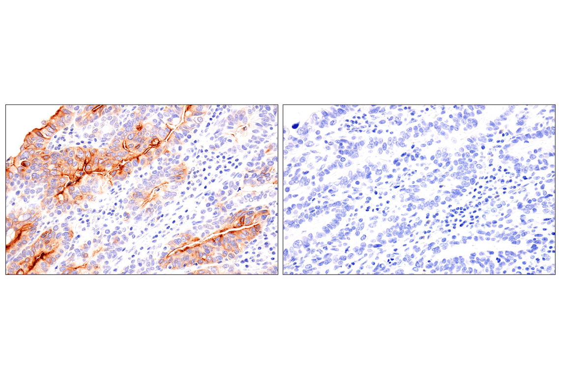
Immunohistochemical analysis of paraffin-embedded OVKATE cell pellet (left, positive) or NCI-H1703 cell pellet (right, negative) using Folate Receptor Alpha/FOLR1 (BN3.2) Mouse mAb.