| Product Includes | Product # | Quantity | Mol. Wt | Isotype/Source |
|---|---|---|---|---|
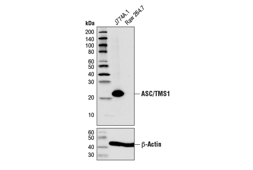
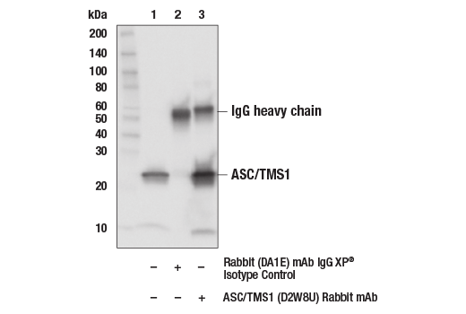
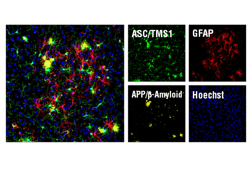
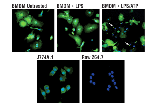
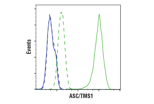
| ASC/TMS1 (D2W8U) Rabbit mAb | 67824 | 20 µl | 22 kDa | Rabbit IgG |
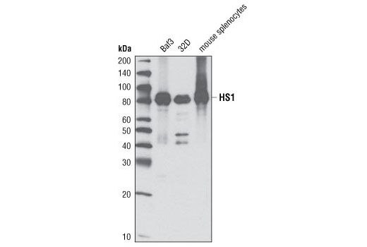
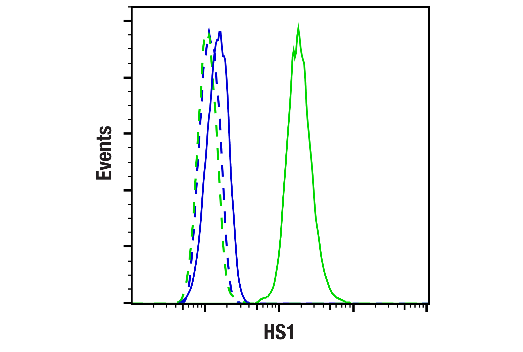
| HS1 (D5A9) XP® Rabbit mAb | 3892 | 20 µl | 80 kDa | Rabbit IgG |
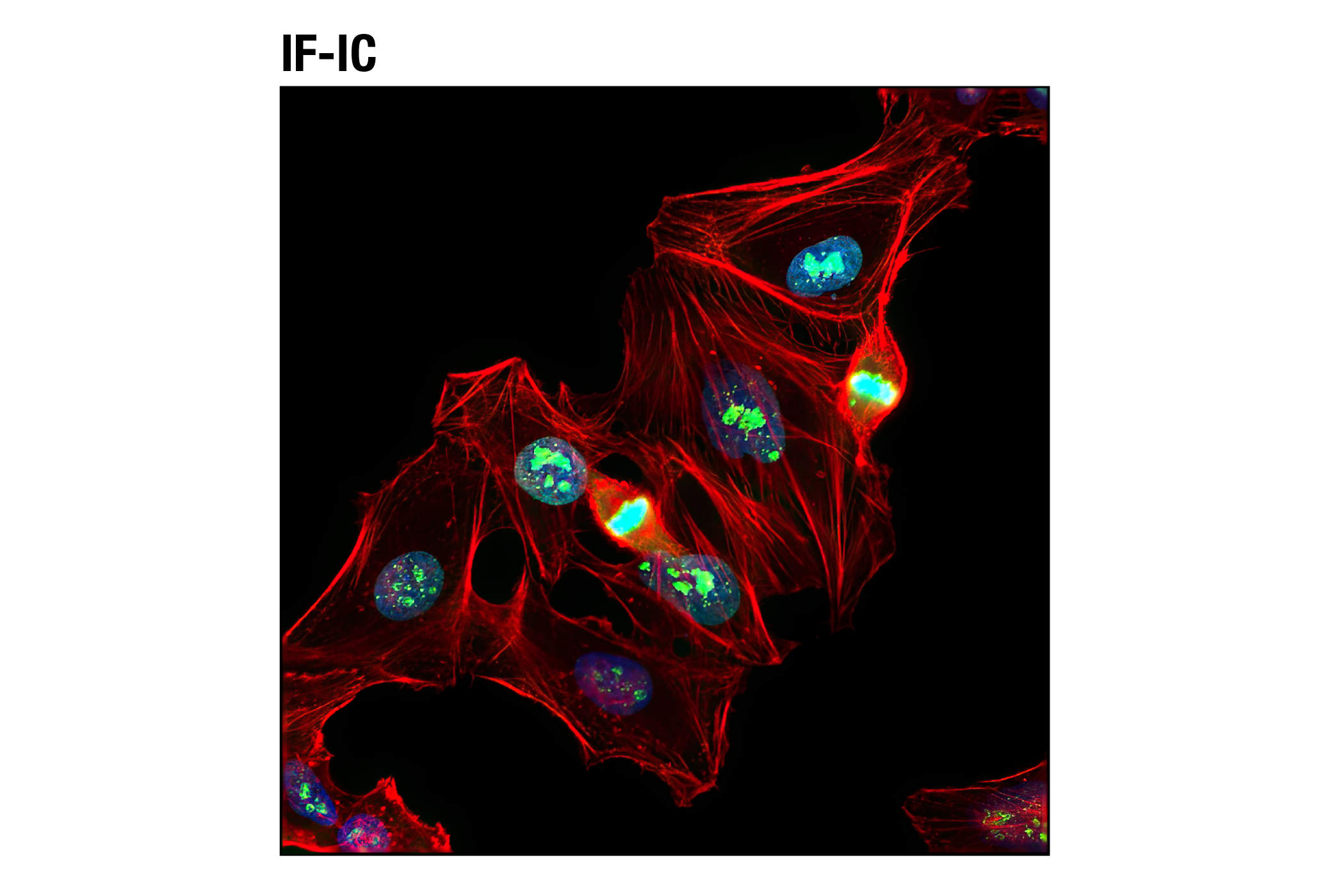
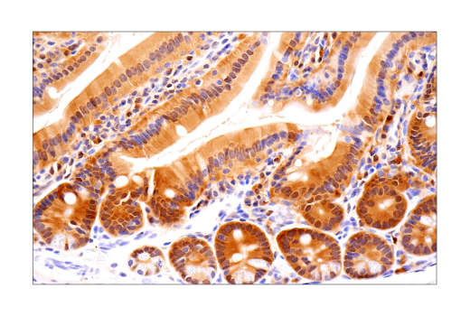
| Ki-67 (D3B5) Rabbit mAb | 9129 | 20 µl | Rabbit IgG | |
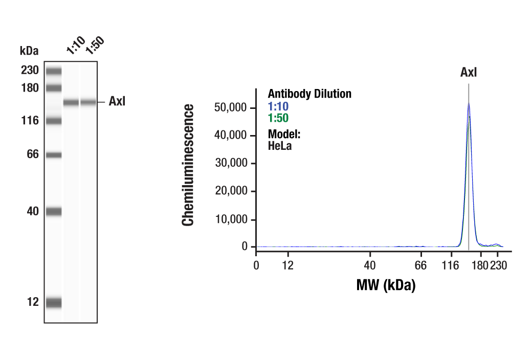
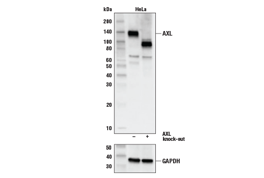
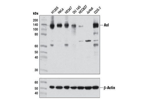
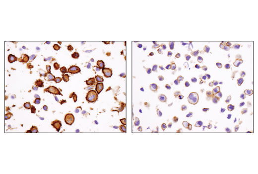
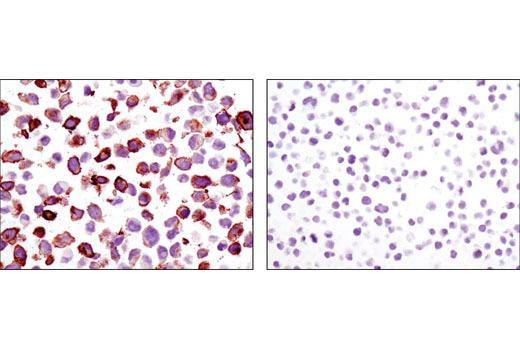
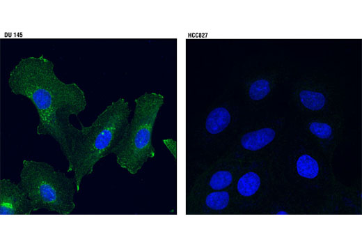
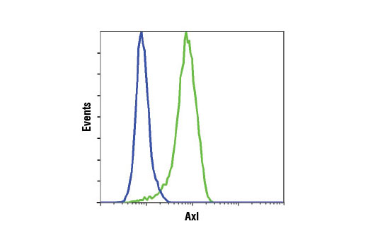
| Axl (C89E7) Rabbit mAb | 8661 | 20 µl | 138 kDa | Rabbit IgG |
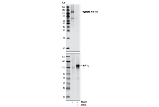
| Hydroxy-HIF-1α (Pro564) (D43B5) XP® Rabbit mAb | 3434 | 20 µl | 120 kDa | Rabbit IgG |
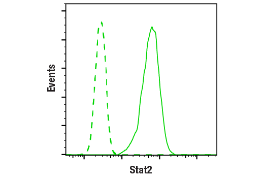
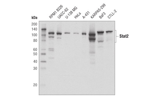
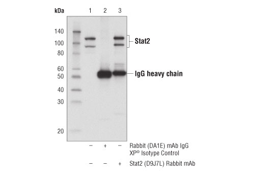
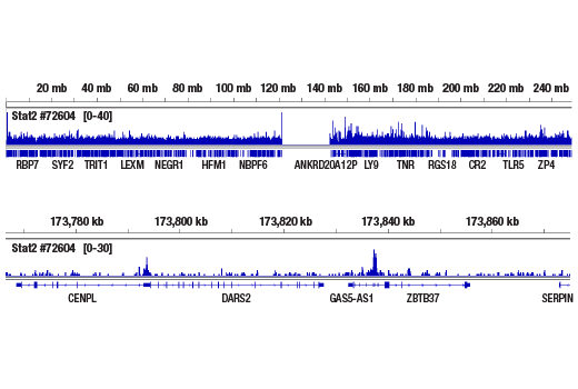
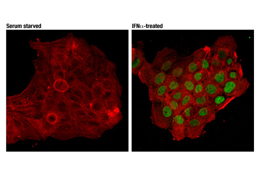
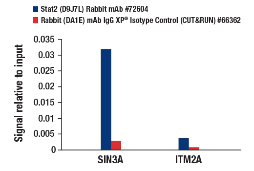
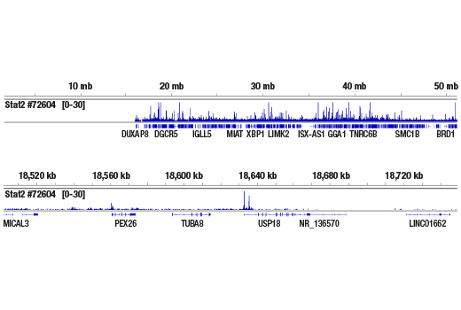
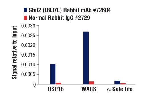
| Stat2 (D9J7L) Rabbit mAb | 72604 | 20 µl | 97, 113 kDa | Rabbit IgG |
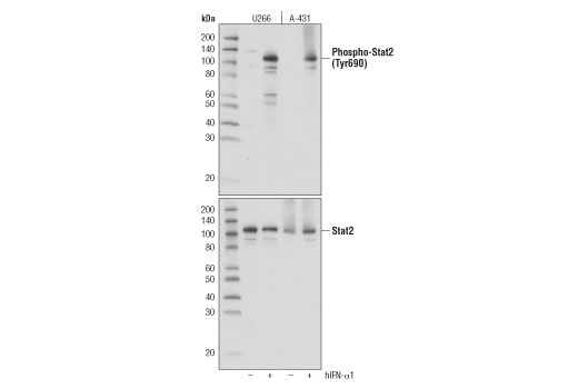
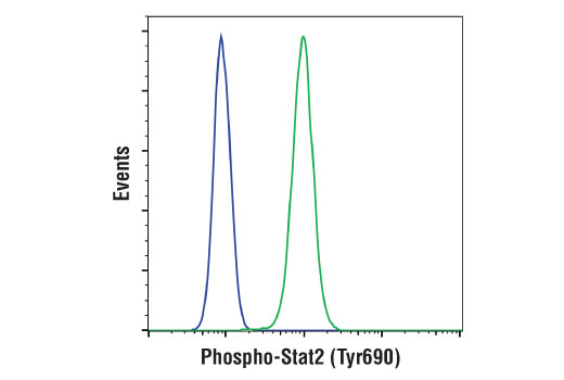
| Phospho-Stat2 (Tyr690) (D3P2P) Rabbit mAb | 88410 | 20 µl | 97, 113 kDa | Rabbit IgG |
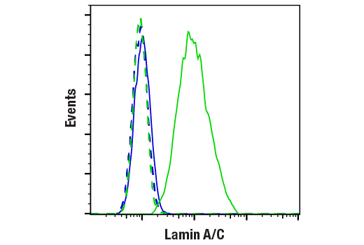
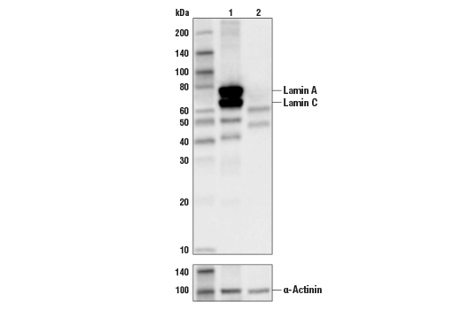
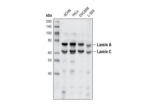
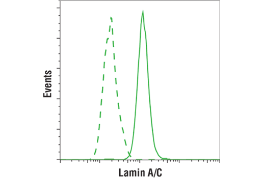
| Lamin A/C (4C11) Mouse mAb | 4777 | 20 µl | 74 (Lamin A), 63 (Lamin C) kDa | Mouse IgG2a |
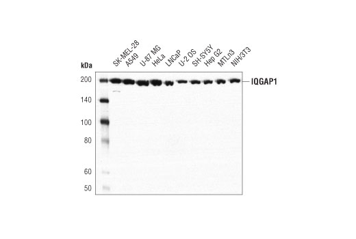
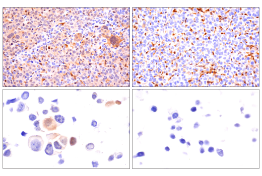
| IQGAP1 (D8K4X) XP® Rabbit mAb | 20648 | 20 µl | 195 kDa | Rabbit IgG |
| Anti-rabbit IgG, HRP-linked Antibody | 7074 | 100 µl | Goat |
Please visit cellsignal.com for individual component applications, species cross-reactivity, dilutions, protocols, and additional product information.
Description
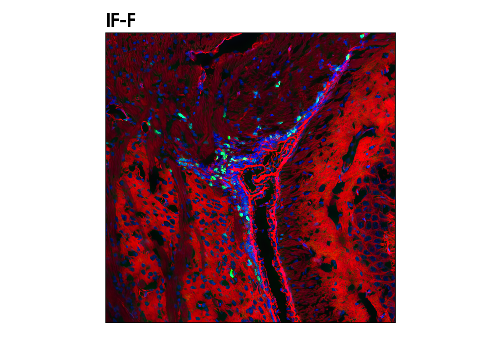
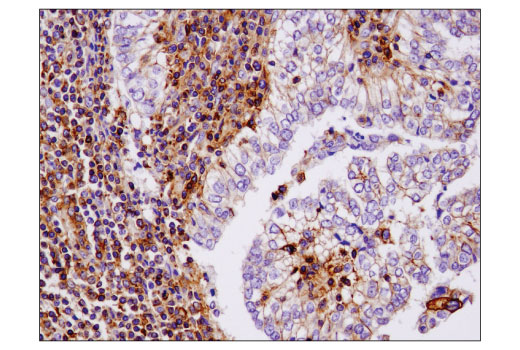
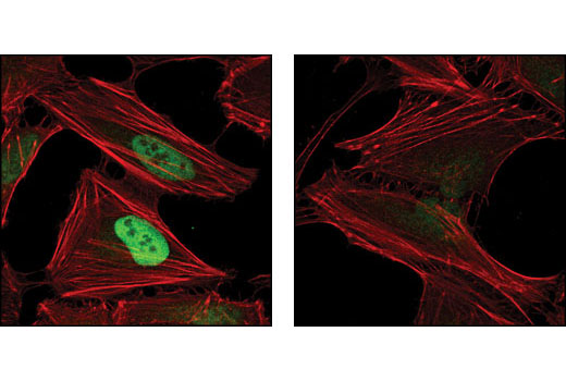
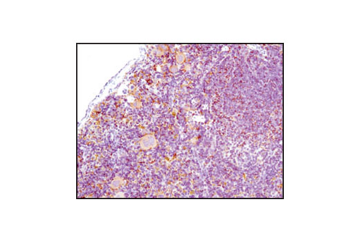
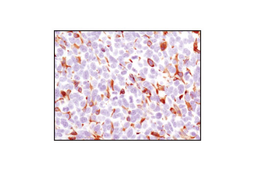
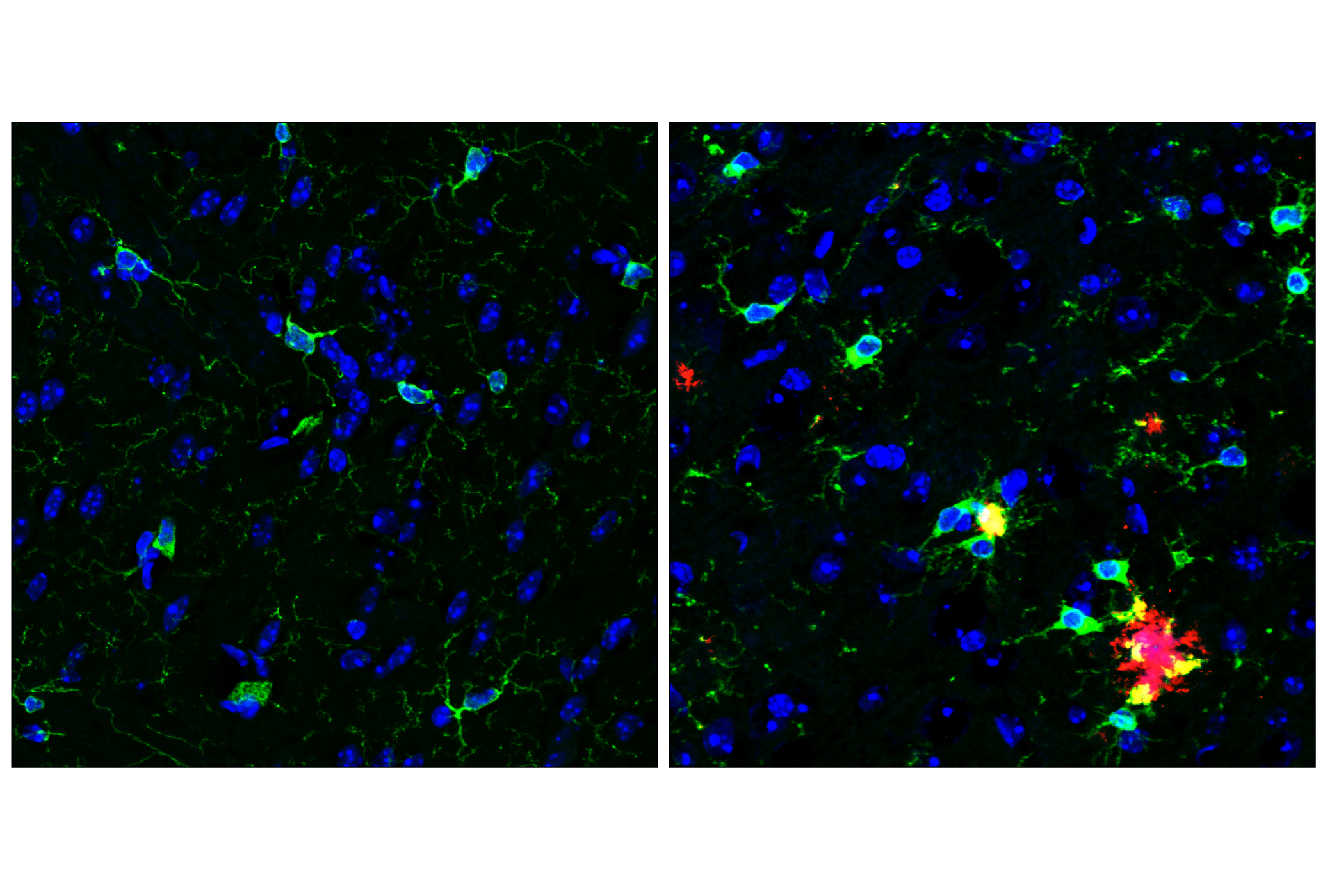
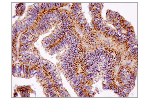
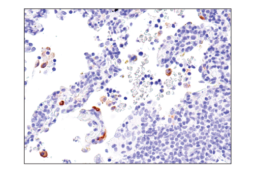
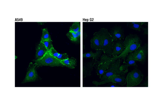
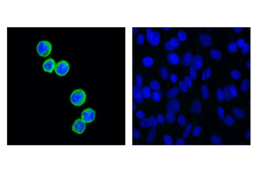
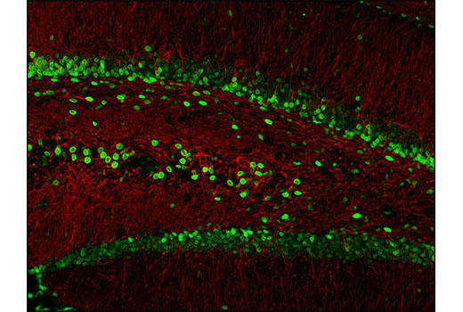
The Microglia Cross Module Antibody Sampler Kit provides an economical means of detecting proteins identified as markers of microglial activity corresponding to proliferation, neurodegeneration, interferon and LPS-relation by western blot and/or immunofluorescence.
Storage
Background
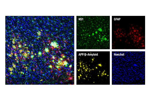
Distinct microglial activation states have been identified using RNA-seq data from a vast array of neurological disease and aging models. These activation states have been categorized into modules corresponding to proliferation, neurodegeneration, interferon-relation, LPS-relation, and many others (1). Previous work identifying markers of specific brain cell types using RNA-seq has shown HS1 and ASC/TMS1 to be useful and specific tools to study microglia (2). HS1 is a protein kinase substrate that is expressed only in tissues and cells of hematopoietic origin (3) and ASC/TMS1 has been found to be a critical component of inflammatory signaling where it associates with and activates caspase-1 in response to pro-inflammatory signals (4).
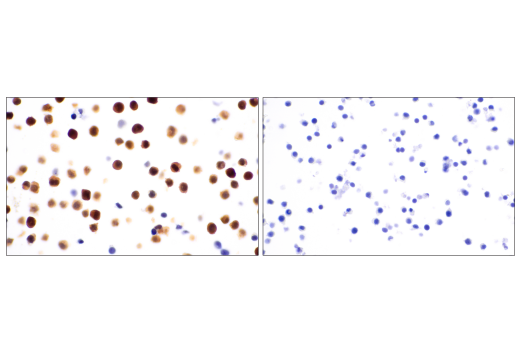
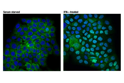
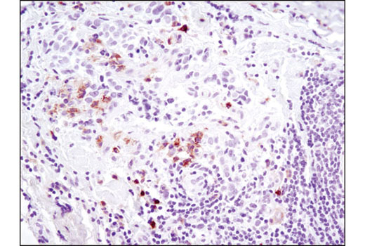
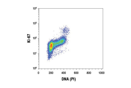
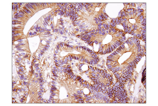
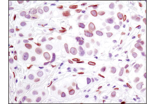
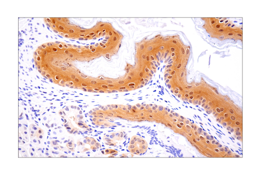
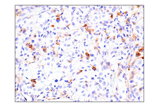
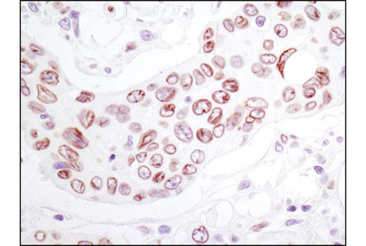
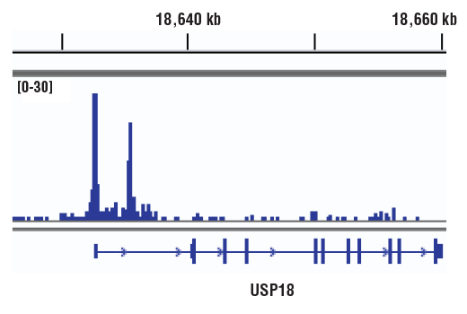
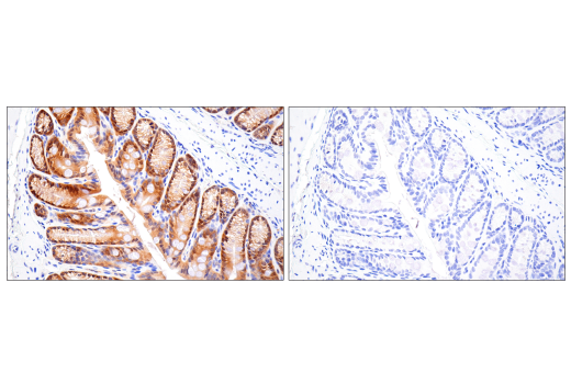
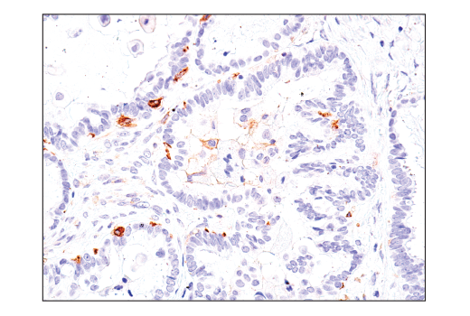
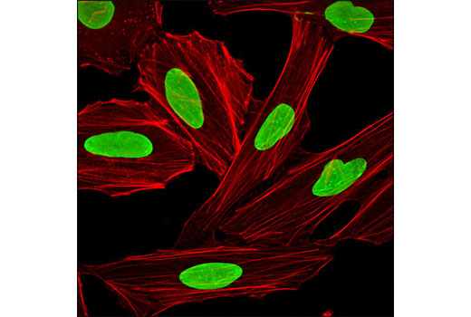
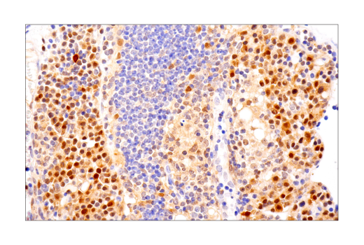
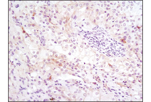
Ki-67 is a nuclear nonhistone protein (5) universally expressed among proliferating cells and absent in quiescent cells (6). Axl is a receptor tyrosine kinase that binds Gas6, stimulating regulatory effects on microglial phagocytic response to inflammatory stimuli (7). Hypoxia inducible factor-1 (HIF-1α) is a transcription factor responsible for adaptation to low oxygen environments whose downstream effects have been shown in a number of neurodegenerative diseases. Under normoxic conditions, HIF-1α is proline hydroxylated leading to ubiquitin mediated degradation (8). Stat2 is critical to the transcriptional responses induced by type I interferons, IFN-alpha/beta (9,10). In response to IFN-alpha/beta, Stat2 is activated by phosphorylation at site Tyr690 through associations with receptor-bound Jak kinases (11). Lamins are nuclear membrane structural components important for maintaining normal cell functions. Lamin A/C is cleaved by caspase-6 and serves as a marker for caspase-6 activation. The cleavage of lamins results in nuclear dysregulation and cell death (12,13). IQGAP1 is ubiquitously expressed and has been found to interact with APC (14) and the CLIP170 complex in response to small GTPases, promoting cell polarization and migration (15).
- Friedman, B.A. et al. (2018) Cell Rep 22, 832-47.
- Zhang, Y. et al. (2014) J Neurosci 34, 11929-47.
- Kitamura, D. et al. (1995) Biochem Biophys Res Commun 208, 1137-46.
- Srinivasula, S.M. et al. (2002) J Biol Chem 277, 21119-22.
- Gerdes, J. et al. (1983) Int J Cancer 31, 13-20.
- Weigel, M.T. and Dowsett, M. (2010) Endocr Relat Cancer 17, R245-62.
- Grommes, C. et al. (2008) J Neuroimmune Pharmacol 3, 130-40.
- Zhang, Z. et al. (2011) Curr Med Chem 18, 4335-43.
- Fu, X.Y. et al. (1992) Proc Natl Acad Sci U S A 89, 7840-3.
- Ihle, J.N. (2001) Curr Opin Cell Biol 13, 211-7.
- Improta, T. et al. (1994) Proc Natl Acad Sci U S A 91, 4776-80.
- Oberhammer, F.A. et al. (1994) J Cell Biol 126, 827-37.
- Rao, L. et al. (1996) J Cell Biol 135, 1441-55.
- Watanabe, T. et al. (2004) Dev Cell 7, 871-83.
- Fukata, M. et al. (2002) Cell 109, 873-85.
Background References
Trademarks and Patents
Limited Uses
Except as otherwise expressly agreed in a writing signed by a legally authorized representative of CST, the following terms apply to Products provided by CST, its affiliates or its distributors. Any Customer's terms and conditions that are in addition to, or different from, those contained herein, unless separately accepted in writing by a legally authorized representative of CST, are rejected and are of no force or effect.
Products are labeled with For Research Use Only or a similar labeling statement and have not been approved, cleared, or licensed by the FDA or other regulatory foreign or domestic entity, for any purpose. Customer shall not use any Product for any diagnostic or therapeutic purpose, or otherwise in any manner that conflicts with its labeling statement. Products sold or licensed by CST are provided for Customer as the end-user and solely for research and development uses. Any use of Product for diagnostic, prophylactic or therapeutic purposes, or any purchase of Product for resale (alone or as a component) or other commercial purpose, requires a separate license from CST. Customer shall (a) not sell, license, loan, donate or otherwise transfer or make available any Product to any third party, whether alone or in combination with other materials, or use the Products to manufacture any commercial products, (b) not copy, modify, reverse engineer, decompile, disassemble or otherwise attempt to discover the underlying structure or technology of the Products, or use the Products for the purpose of developing any products or services that would compete with CST products or services, (c) not alter or remove from the Products any trademarks, trade names, logos, patent or copyright notices or markings, (d) use the Products solely in accordance with CST Product Terms of Sale and any applicable documentation, and (e) comply with any license, terms of service or similar agreement with respect to any third party products or services used by Customer in connection with the Products.