

High-Content Imaging
Leveraging High-Content Screening & High-Content Analysis
To achieve high-content imaging (HCI) success, you need to rely on high-quality, trustworthy reagents. CST has high-content analysis (HCA)/high-content screening (HCS) antibodies you can trust to work consistently on your HCI instrument. Use CST® antibodies with confidence to accelerate your screening assays.
- Thousands of CST antibodies are available for high-content imaging.
- All CST antibodies approved for immunofluorescence (IF) have been tested and shown to perform well on HCA instruments.
- Save time and resources using high-quality CST antibodies.
Fluorescent Conjugates and Custom Conjugation
Many IF-approved CST antibodies, which are ideal for high-content imaging, are available as fully validated fluorescent antibody conjugates. Can’t find the conjugated antibody that you’re looking for? We also offer custom conjugation services to simplify your workload and streamline your project.
Why choose IF/HCA-validated CST antibodies?
Advantages
CST antibodies are developed with unparalleled validation testing, providing reproducible results for your high-content imaging assay. Expert CST scientists validate every immunofluorescence antibody in-house in multiple ways, including in high-content analysis assays. Learn more about our extensive validation testing.
Thousands of Antibodies for High-Content Imaging
Our catalog of IF-approved antibodies continues to grow every day. These HCA/HCS-ready antibodies are ideal for examining:
- Critical disease signaling pathways
- Protein target expression
- Post-translational protein modifications
- Subcellular localization
CST antibodies can help you obtain robust fluorescence microscopy images while providing quantitative endpoint analysis for your disease modeling studies. Rest assured, our antibodies will perform optimally as you screen molecular candidates in your high-content analysis or high-content screening assays.
Bulk Orders, Lot Reservations, and Custom Formulations
All CST antibody products can be purchased in bulk quantities or with antibody lot reservations. To meet your needs, they can also be provided in alternative formulations without BSA or sodium azide. Contact us for more information.
Discover the Power of High-Content Imaging
High-Content Imaging Research Examples
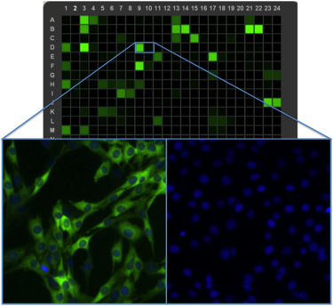
Automated techniques for high-content imaging, like high-content analysis and high-content screening, offer great promise for gaining a deeper understanding of cellular mechanisms in both normal and disease states. These methods can provide an efficient way to evaluate biological processes on multiple levels while providing additional benefits such as increased efficiency and reduced intrinsic user bias.
However, it is crucial to obtain reliable, reproducible, and statistically robust data from high-throughput experiments, so it’s essential to choose your antibodies and reagents carefully. The following videos, posters, and application notes demonstrate how high-content imaging can be used to evaluate biological systems and pathways.
Find out more about how to optimize your high-throughput, high-content analysis, and high-content screening assays using the following detailed CST Application Notes.
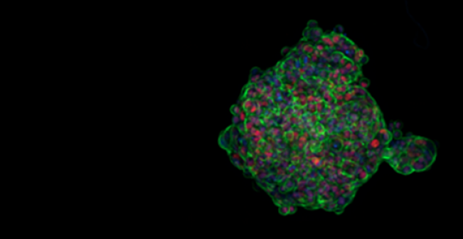
3D/Organoid Imaging Using CST IF/HCS Antibodies
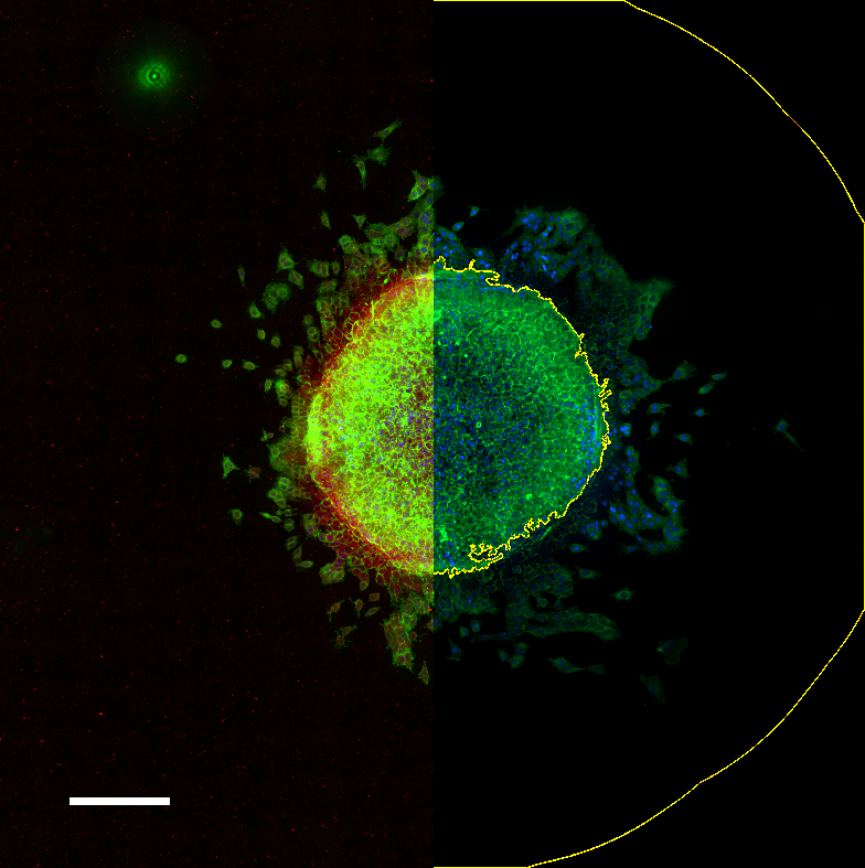
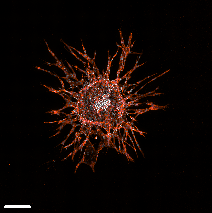
Imaged with the confocal imaging mode of the Agilent Cytation C10 (which enables optical resolution of Z-planes in 3D biological samples).
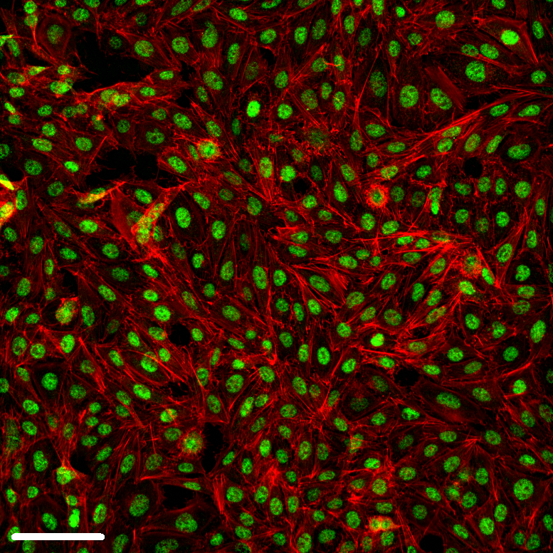
Cell Painting Using CST IF/HCS Antibodies
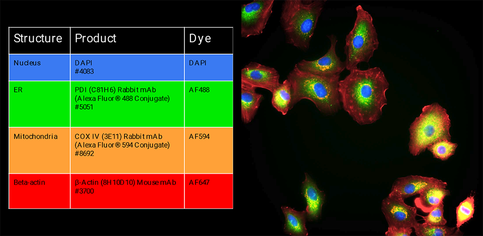
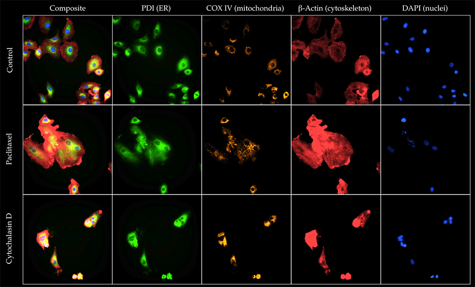
Images obtained using the Operetta CLS High-Content Analysis System made by Revvity.