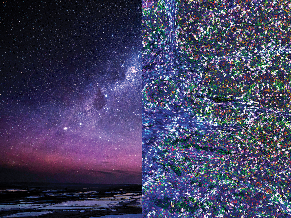
SignalStar Multiplex Immunohistochemistry (mIHC) is a methodology for studying FFPE tissue samples. It uses antibodies, oligonucleotides, and fluorophores to identify which cells are present, where they are located, and what functions they perform.
SignalStar technology enables the detection of multiple phenotypic and functional biomarkers while preserving spatial context and tissue architecture. These insights are critical for understanding how cells organize and interact to influence the tissue microenvironment that ultimately drives disease progression and response to therapy.
The resources we've put together below can provide support for integrating SignalStar technology into your spatial biology research.
The SignalStar Multiplex IHC Panel Builder provides automated panel design, pairing antibodies to fluorophores and imaging rounds with efficient accuracy. Panel builder results can be edited for complete control of design. If you choose to modify your panel, keep these considerations in mind:
Protein Abundance: Channels 594 and 647 are brightest, and should be paired with antibodies binding the least abundant antigens. Channel 750 has a lower signal intensity, and the panel builder will prohibit some antibodies from pairing with the 750 channel.
Autofluorescence: Antibodies expected to detect low abundance proteins in your tissue should be in channels less affected by autofluorescence. Channel 488 is most impacted by autofluorescence and should be avoided for these biomarkers.
Imaging Rounds: While the majority of antibodies should be strong and clean regardless of imaging round, signal intensity may be impacted by low target expression levels and poor tissue quality. For best results, antibodies against lower abundance proteins should be placed in the first imaging round, while antibodies against higher abundance markers should be placed in the second imaging round. The panel builder may prohibit some antibodies from being used in certain imaging rounds.
Imaging Instrumentation: To avoid spectral bleed-through, consider the spectral characteristics of your fluorescence microscope. Epifluorescent, confocal, and scanning fluorescence microscopes can typically distinguish between the SignalStar channels:
Fluorophore Channel | Excitation (nm) | Emission (nm) | Laser Line | Common Filter Set |
488 | 488 | 520 | 488 | FITC |
594 | 590 | 618 | 561/594 | Texas Red |
647 | 650 | 668 | 594/633 | Cy®5 |
750 | 752 | 776 | 633 | Cy®7 |
If your final panel includes all four (4) fluorescent channels, in addition to DAPI, then you'll need to confirm your fluorescent imaging instrumentation can specifically detect and separate these channels based on these parameters.
Spectral Unmixing: The antibodies, reagents, and protocols included in your SignalStar Multiplex IHC Panel are optimized to minimize spectral overlap. To further ensure that fluorescence from individual antibodies can be differentiated, we recommend creating a spectral library using slides stained individually with the channels described above.
If the characteristics of your imaging instrument are not ideal for separating these fluorescent channels, a spectral library can be used to computationally unmix fluorescent signals.
Cell Signaling Technology, CST, and SignalStar are trademarks of Cell Signaling Technology, Inc. Cy is a registered trademark of GE Healthcare. All other trademarks are the property of their respective owners. Visit cellsignal.com/trademarks for more information.
U.S. Patent No. 10,781,477, foreign equivalents, and child patents deriving therefrom.