SignalStar™ mIHC Panel Design
Before You Get Started
SignalStar Multiplex IHC technology uses specific primary antibodies and fluorescent detection reagents to label cellular proteins in FFPE tissue samples. It enables you to quantify multiple biomarkers simultaneously using highly-validated antibody panels for spatial biology experiments.
The SignalStar Multiplex IHC Panel Builder automates your panel design, eliminating the guesswork of pairing antibodies to fluorophores to ensure accuracy. Experimental design is done in four basic steps, allowing you to quickly and easily choose your target species, antibodies, fluorophores, and imaging sequence. Or you can choose your species and antibodies, and let the automated panel builder guide you the rest of the way.
Confirm Your Instrument Compatibility
- SignalStar Multiplex IHC protocols can be performed either manually at your lab bench or automated on the BOND RX autostainer by Leica Biosystems. See the protocols for more details regarding instrumentation requirements.
- Consider the spectral characteristics of your fluorescent imaging instrumentation to achieve maximum results and avoid spectral bleed-through.
- If your final panel includes all four (4) fluorescent channels, in addition to DAPI, then you'll need to confirm your fluorescent imaging instrumentation can specifically detect and separate these channels based on the parameters below:
Fluorophore Channel | Excitation (nm) | Emission (nm) | Laser Line | Common Filter Set |
488 | 488 | 520 | 488 | FITC |
594 | 590 | 618 | 561/594 | Texas Red |
647 | 650 | 668 | 594/633 | Cy®5 |
750 | 752 | 776 | 633 | Cy®7 |
How to Design Your SignalStar Multiplex IHC Panel
Step 1: Choose Your Target Species
SignalStar Multiplex IHC antibodies are validated to confirm specific reactivity with the species indicated. Not all antibodies work in all species.
Step 2: Choose Your Antibodies
Choose 3-8 antibodies validated for your markers of interest. Like all CST® antibodies, the ones identified for use in your SignalStar panels have each been individually validated for use in their intended application. So they're guaranteed to perform as expected, and to provide you with reliable, reproducible results.
Step 3: View and Edit Your Panel Builder Results
The panel builder automates your mIHC panel design, pairing antibodies to fluorophores and imaging rounds with inherent accuracy and efficiency. You can review and confirm the panel builder results, or choose to edit for more control of your experimental design.
Considerations when designing your own Multiplex IHC Panel
Step 4: Confirm Your Order
All antibodies and buffer kits needed for your experiment are automatically added to your cart. You can continue to add panels in the panel builder multiple times. Just be careful not to remove components of prior panels once they're in your basket.
The SignalStar mIHC Workflow
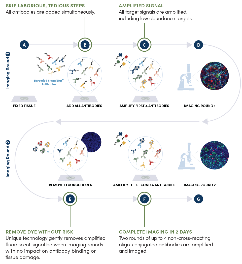
The SignalStar mIHC assay allows for the simultaneous labeling of up to 8 biomarkers in FFPE tissue. Deparaffinized and rehydrated FFPE tissue sections undergo antigen retrieval (A), and all antibodies in your plex size of choice (3-8 maximum unique oligo-conjugated antibodies) are added in a cocktail, in one primary incubation step (B). A network of complementary oligos with fluorescent dyes (channels: 488, 594, 647, 750) amplify the signal of up to 4 oligo-conjugated antibodies in the first round of imaging by building oligo-fluorophore constructs attached to the antibody (C-D). If the plex size is greater than 4, the first round of oligonucleotides and fluorophores are gently removed (E), and a second round of amplification is performed to visualize up to 4 additional oligo-conjugated antibodies (F). The two images are then aligned and fused computationally with either proprietary or open-source software to generate the 8-plex image (G).
Need Support?
CST provides expert support to help you optimize the use of SignalStar Multiplex IHC in your research.
Visit the Technical Support page to search for SignalStar-related troubleshooting information and answers to technical questions.
Cell Signaling Technology, CST, and SignalStar are trademarks of Cell Signaling Technology, Inc. Cy is a registered trademark of GE Healthcare. All other trademarks are the property of their respective owners. Visit /legal/trademark-information for more information.
U.S. Patent No. 10,781,477, foreign equivalents, and child patents deriving therefrom.


