

Multiplex IHC
Our extensive validation and advanced technical support ensures that all of our IHC-P-validated antibodies can be used for fluorescent multiplex IHC.
Benefits of multiplex IHC
- Gather maximal data per tissue section, which is critical when tissue samples are limited.
- Understand co-expression and spatial organization of multiple targets within preserved tissue architecture, unlike alternative multiplex approaches (e.g., next generation sequencing, PCR, mass spectrometry, etc.
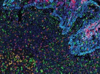
Being able to visualize multiple targets simultaneously is important in research fields like tumor immunology where it is necessary to catalog subsets of immune and cancer cells within the tumor microenvironment. For example, tumors may evade immune detection by manipulating the expression of immune checkpoint proteins (e.g., PD-1, CTLA-4, TIM-3) to “turn off” activated T cells. Alternatively, T cells may become less effective at destroying tumor cells as they succumb to exhaustion, a phenomenon characterized by changes in immune checkpoint protein expression, T cell expression of VISTA, and the appearance of CD68+ co-infiltrating macrophages. Thus, molecular profiling of immune checkpoint proteins together with immune cell phenotyping is key to understanding the complex tumor microenvironment and to enabling the development of tailored, combinatorial therapeutic intervention.
Multiplexing options
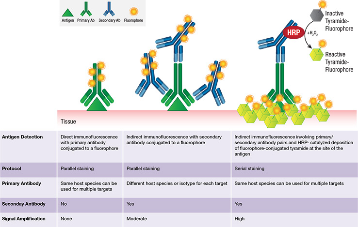
Benefits of tyramide-based multiplexing:
- Signal amplification:
- Provides marked enhancement of antigen-associated fluorescence signal.
- Enables detection of low abundance targets.
- Simplified panel design: Compatible with any IHC-P validated antibody regardless of the host species or isotype.