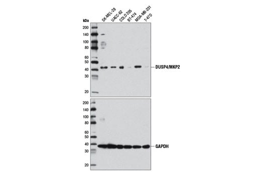
DUSP4/MKP2 (D9A5) Rabbit mAb #5149
- WB
To Purchase # 5149
| Cat. # | Size | Qty. | Price | Ships |
|---|---|---|---|---|
| 5149T | 20 µl | $188 | ||
| 5149S | 100 µl | $399 |
Supporting Data
| REACTIVITY | H Mk |
| SENSITIVITY | Endogenous |
| MW (kDa) | 42 |
| Source/Isotype | Rabbit |
Application Key:
- WB-Western Blotting
Species Cross-Reactivity Key:
- H-Human
- Mk-Monkey
- Related Products
Product Information
Product Usage Information
| Application | Dilution |
|---|---|
| Western Blotting | 1:1000 |
| Simple Western™ | 1:50 - 1:250 |
Storage
Protocol
Specificity / Sensitivity
Species Reactivity:
Source / Purification
Background
DUSP4 (MKP2, hVH2) is a nuclear dual-specificity phosphatase that is a negative regulator of Erk1/2 signaling by dephosphorylating and inactivating Erk1/2 in response to growth factors (7,8). Treatment with mitogen or expression of activating mutations of Ras (G12V) or Raf (V600E) promote increased expression of DUSP4 and a coincident decrease in phospho-Erk in the nucleus (9). In contrast, numerous studies have detected decreased expression of DUSP4 in a variety of tumor types, resulting in increased signaling via the Ras/Erk pathway, enhanced tumor growth, and decreased drug sensitivity (10-12). DUSP4/MKP2 also plays an important role in regulating the immune system where it has been implicated in regulating T and B cell proliferation and apoptosis, and adaptive and inflammatory responses (13-16).
- Camps, M. et al. (2000) FASEB J 14, 6-16.
- Theodosiou, A. and Ashworth, A. (2002) Genome Biol 3, REVIEWS3009.
- Salojin, K. and Oravecz, T. (2007) J Leukoc Biol 81, 860-9.
- Tanoue, T. et al. (2002) J Biol Chem 277, 22942-9.
- Dickinson, R.J. and Keyse, S.M. (2006) J Cell Sci 119, 4607-15.
- Wu, G.S. (2007) Cancer Metastasis Rev 26, 579-85.
- Peng, D.J. et al. (2010) Cell Cycle 9, 4650-5.
- Lawan, A. et al. (2011) J Biol Chem 286, 12933-43.
- Cagnol, S. and Rivard, N. (2012) Oncogene , .
- Chitale, D. et al. (2009) Oncogene 28, 2773-83.
- Waha, A. et al. (2010) Cancer Res 70, 1689-99.
- Balko, J.M. et al. (2012) Nat Med 18, 1052-9.
- Ramesh, S. et al. (2008) EMBO Rep 9, 990-7.
- Cornell, T.T. et al. (2010) Infect Immun 78, 2868-76.
- Huang, C.Y. et al. (2012) Eur J Immunol 42, 476-88.
- Yu, M. et al. (2012) Proc Natl Acad Sci U S A 109, E879-88.
Pathways
Explore pathways related to this product.
Limited Uses
Except as otherwise expressly agreed in a writing signed by a legally authorized representative of CST, the following terms apply to Products provided by CST, its affiliates or its distributors. Any Customer's terms and conditions that are in addition to, or different from, those contained herein, unless separately accepted in writing by a legally authorized representative of CST, are rejected and are of no force or effect.
Products are labeled with For Research Use Only or a similar labeling statement and have not been approved, cleared, or licensed by the FDA or other regulatory foreign or domestic entity, for any purpose. Customer shall not use any Product for any diagnostic or therapeutic purpose, or otherwise in any manner that conflicts with its labeling statement. Products sold or licensed by CST are provided for Customer as the end-user and solely for research and development uses. Any use of Product for diagnostic, prophylactic or therapeutic purposes, or any purchase of Product for resale (alone or as a component) or other commercial purpose, requires a separate license from CST. Customer shall (a) not sell, license, loan, donate or otherwise transfer or make available any Product to any third party, whether alone or in combination with other materials, or use the Products to manufacture any commercial products, (b) not copy, modify, reverse engineer, decompile, disassemble or otherwise attempt to discover the underlying structure or technology of the Products, or use the Products for the purpose of developing any products or services that would compete with CST products or services, (c) not alter or remove from the Products any trademarks, trade names, logos, patent or copyright notices or markings, (d) use the Products solely in accordance with CST Product Terms of Sale and any applicable documentation, and (e) comply with any license, terms of service or similar agreement with respect to any third party products or services used by Customer in connection with the Products.


