

RIP3 (E7A7F) XP® Rabbit mAb (Alexa Fluor® 647 Conjugate) #36718
- F
To Purchase # 36718
| Cat. # | Size | Qty. | Price |
|---|---|---|---|
| 36718S | 100 µl 50 tests | $450 |
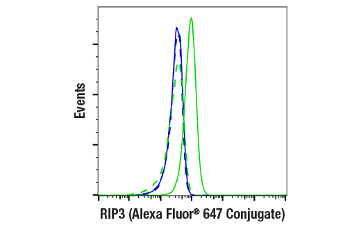
Supporting Data
| REACTIVITY | H |
| SENSITIVITY | Endogenous |
| MW (kDa) | |
| Source/Isotype | Rabbit IgG |
Application Key:
- F-Flow Cytometry
Species Cross-Reactivity Key:
- H-Human
- Related Products
Product Information
Product Description
Product Usage Information
| Application | Dilution |
|---|---|
| Flow Cytometry (Fixed/Permeabilized) | 1:50 |
Storage
Protocol
Specificity / Sensitivity
Species Reactivity:
Source / Purification
Background
Receptor-interacting protein 3 (RIP3) was originally found to interact with RIP and the TNF receptor complex to induce apoptosis and activation of NF-κB (9,10). It has subsequently been shown that the association between RIP and RIP3 is a key component of a signaling pathway that results in programmed necrosis (necroptosis), a necrotic-like cell death induced by TNF in the presence of caspase inhibitors (11-13). RIP3 is phosphorylated at Ser227 and targets the phosphorylation of mixed lineage kinase domain-like protein (MLKL), which is critical for necroptosis (14).
- Meylan, E. and Tschopp, J. (2005) Trends Biochem Sci 30, 151-9.
- Hsu, H. et al. (1996) Immunity 4, 387-96.
- Stanger, B.Z. et al. (1995) Cell 81, 513-23.
- Ting, A.T. et al. (1996) EMBO J 15, 6189-96.
- Kelliher, M.A. et al. (1998) Immunity 8, 297-303.
- Devin, A. et al. (2000) Immunity 12, 419-29.
- Zhang, S.Q. et al. (2000) Immunity 12, 301-11.
- Lin, Y. et al. (1999) Genes Dev 13, 2514-26.
- Yu, P.W. et al. (1999) Curr Biol 9, 539-42.
- Sun, X. et al. (1999) J Biol Chem 274, 16871-5.
- Zhang, D.W. et al. (2009) Science 325, 332-6.
- He, S. et al. (2009) Cell 137, 1100-11.
- Cho, Y.S. et al. (2009) Cell 137, 1112-23.
- Sun, L. et al. (2012) Cell 148, 213-27.
Pathways
Explore pathways related to this product.
Limited Uses
Except as otherwise expressly agreed in a writing signed by a legally authorized representative of CST, the following terms apply to Products provided by CST, its affiliates or its distributors. Any Customer's terms and conditions that are in addition to, or different from, those contained herein, unless separately accepted in writing by a legally authorized representative of CST, are rejected and are of no force or effect.
Products are labeled with For Research Use Only or a similar labeling statement and have not been approved, cleared, or licensed by the FDA or other regulatory foreign or domestic entity, for any purpose. Customer shall not use any Product for any diagnostic or therapeutic purpose, or otherwise in any manner that conflicts with its labeling statement. Products sold or licensed by CST are provided for Customer as the end-user and solely for research and development uses. Any use of Product for diagnostic, prophylactic or therapeutic purposes, or any purchase of Product for resale (alone or as a component) or other commercial purpose, requires a separate license from CST. Customer shall (a) not sell, license, loan, donate or otherwise transfer or make available any Product to any third party, whether alone or in combination with other materials, or use the Products to manufacture any commercial products, (b) not copy, modify, reverse engineer, decompile, disassemble or otherwise attempt to discover the underlying structure or technology of the Products, or use the Products for the purpose of developing any products or services that would compete with CST products or services, (c) not alter or remove from the Products any trademarks, trade names, logos, patent or copyright notices or markings, (d) use the Products solely in accordance with CST Product Terms of Sale and any applicable documentation, and (e) comply with any license, terms of service or similar agreement with respect to any third party products or services used by Customer in connection with the Products.