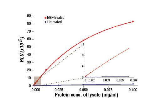
PathScan® Phospho-EGF Receptor (Tyr1068) Chemiluminescent Sandwich ELISA Kit #7036
- ELISA
To Purchase # 7036
| Cat. # | Size | Qty. | Price |
|---|---|---|---|
| 7036C | 1 Kit 96 assays | $641 | |
| 7036V1 | 5 Kits 480 assays | $3,125 | |
| 7036V2 | 10 Kits 960 assays | $6,090 | |
| 7036V3 | 20 Kits 1920 assays | $11,859 | |
| 7036V4 | 50 Kits 4800 assays | $28,845 |
Important Ordering Details
Custom Ordering Details:If kit quantities from the same lot are needed in unlisted sizes, contact us for processing time and pricing.
Looking for this ELISA kit in a 384-well format? Inquire for availability, processing time, and pricing.
Supporting Data
| REACTIVITY | H |
Application Key:
- ELISA-ELISA
Species Cross-Reactivity Key:
- H-Human
- Product Includes
- Related Products
| Product Includes | Volume | Solution Color |
|---|---|---|
| EGF Receptor Mouse mAb Coated Microwells #99592 | 96 tests | |
| Phospho-EGF Receptor (Tyr1068) Rabbit Detection mAb #13019 | 1 ea | Green (Lyophilized) |
| Anti-rabbit IgG, HRP-linked Antibody (ELISA Formulated) #13272 | 1 ea | Red (Lyophilized) |
| Detection Antibody Diluent #13339 | 5.5 ml | Green |
| HRP Diluent #13515 | 5.5 ml | Red |
| Luminol/Enhancer Solution #84850 | 3 ml | |
| Stable Peroxide Buffer #42552 | 3 ml | |
| Sealing Tape #54503 | 2 ea | |
| ELISA Wash Buffer (20X) #9801 | 25 ml | |
| ELISA Sample Diluent #11083 | 25 ml | Blue |
| Cell Lysis Buffer (10X) #9803 | 15 ml |
Kit contents scale proportionally with size, except sealing tape.
Example: The V1 kit contains 5X the listed quantities above, but will exclude the sealing tape.
The microwell plate is supplied as 12 8-well modules - Each module is designed to break apart for 8 tests.
Product Information
Product Description
*Antibodies in this kit are custom formulations specific to kit.
Protocol
Specificity / Sensitivity
Species Reactivity:
Background
- Hackel, P.O. et al. (1999) Curr Opin Cell Biol 11, 184-9.
- Zwick, E. et al. (1999) Trends Pharmacol Sci 20, 408-12.
- Cooper, J.A. and Howell, B. (1993) Cell 73, 1051-4.
- Hubbard, S.R. et al. (1994) Nature 372, 746-54.
- Biscardi, J.S. et al. (1999) J Biol Chem 274, 8335-43.
- Emlet, D.R. et al. (1997) J Biol Chem 272, 4079-86.
- Levkowitz, G. et al. (1999) Mol Cell 4, 1029-40.
- Ettenberg, S.A. et al. (1999) Oncogene 18, 1855-66.
- Rojas, M. et al. (1996) J Biol Chem 271, 27456-61.
- Feinmesser, R.L. et al. (1999) J Biol Chem 274, 16168-73.
Pathways
Explore pathways related to this product.
Limited Uses
Except as otherwise expressly agreed in a writing signed by a legally authorized representative of CST, the following terms apply to Products provided by CST, its affiliates or its distributors. Any Customer's terms and conditions that are in addition to, or different from, those contained herein, unless separately accepted in writing by a legally authorized representative of CST, are rejected and are of no force or effect.
Products are labeled with For Research Use Only or a similar labeling statement and have not been approved, cleared, or licensed by the FDA or other regulatory foreign or domestic entity, for any purpose. Customer shall not use any Product for any diagnostic or therapeutic purpose, or otherwise in any manner that conflicts with its labeling statement. Products sold or licensed by CST are provided for Customer as the end-user and solely for research and development uses. Any use of Product for diagnostic, prophylactic or therapeutic purposes, or any purchase of Product for resale (alone or as a component) or other commercial purpose, requires a separate license from CST. Customer shall (a) not sell, license, loan, donate or otherwise transfer or make available any Product to any third party, whether alone or in combination with other materials, or use the Products to manufacture any commercial products, (b) not copy, modify, reverse engineer, decompile, disassemble or otherwise attempt to discover the underlying structure or technology of the Products, or use the Products for the purpose of developing any products or services that would compete with CST products or services, (c) not alter or remove from the Products any trademarks, trade names, logos, patent or copyright notices or markings, (d) use the Products solely in accordance with CST Product Terms of Sale and any applicable documentation, and (e) comply with any license, terms of service or similar agreement with respect to any third party products or services used by Customer in connection with the Products.


