

PD-L1
The immune system employs a series of checkpoints to protect normal, healthy tissue from an immune response. These consist of receptors on the surface of activated T cells and their corresponding ligands on the surface of antigen presenting cells. A key immune checkpoint is triggered when PD-1 (programmed cell death protein 1) engages its ligand PD-L1. As a result of this interaction, T cell activation is attenuated and an active immune response is prevented(1).
This mechanism is often co-opted by tumors. PD-L1 is upregulated in several tumor types and contributes to the malignancy of these cancers by interacting with PD-1 and inhibiting T cell activation. In this way, the tumors avoid detection and destruction by the immune system(1-3). Accordingly, PD-1 and PD-L1 have garnered much attention for their roles in tumor immunology and as immune-based therapeutic targets(4,5).
CST PD-L1 product comparison for IHC-P analysis
| Product Name | PD-L1 (E1L3N®) XP® Rabbit mAb #13684 | PD-L1 (405.9A11) Mouse mAb #29122 | PD-L1 (Extracellular Domain Specific) (E1J2J) Rabbit mAb #15165 |
|---|---|---|---|
| Clone | E1L3N® | 405.9A11 | E1J2J |
| Epitope | Intracellular | Intracellular | Extracellular |
| Antibody host species | Rabbit | Mouse | Rabbit |
| IHC analysis of paraffin-embedded human placenta | 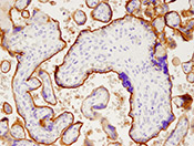 |
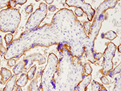 |
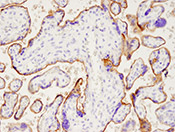 |
| Recommended usage for IHC | Preferred clone based on sensitivity | Use when multiplexing with rabbit antibodies | Use for studying the extracellular domain of PD-L1 |