Neurons are CNS cells that receive and transmit electrochemical signals by a process called neurotransmission. The anatomy of the neurons is conducive to the receipt and dispensing of information: axons send signals to neurons and dendrites receive signals from other neurons.
A neuron is categorized as excitatory or inhibitory based on the type of signal it releases. These signals can hyperpolarize or depolarize their target neurons. Other characteristics that contribute to the classification of the neuron include its polarity, morphology, anatomical location, protein expression profile, and directional flow of information.
Neurotransmission from the neuron and extraneural systems moves through a series of intracellular events to mediate the propagation of the electrical signal. The extracellular space between two cells is the synapse, and therefore the signal origin is the presynaptic cell, and the receiving neuron is the postsynaptic cell. Neurotransmitters are signaling molecules that generate an excitatory or inhibitory response on the postsynaptic membrane, thereby propagating or preventing an action potential.
Neurotransmitters can be categorized as small molecule or neuropeptides. The synthesis of small molecule neurotransmitters occurs locally - within the axon terminal, whereas neuropeptides are much larger than small molecules, and therefore are synthesized within the cell body.

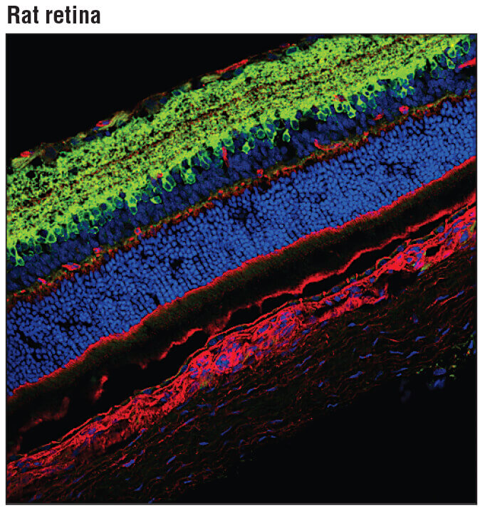
Confocal immunofluorescent analysis of rat retina using GAD1 (A9A5X) Rabbit mAb (green). Actin filaments were labeled with DyLight 554 Phalloidin #13054 (red). Blue pseudocolor = DRAQ5 #4084 (fluorescent DNA dye).

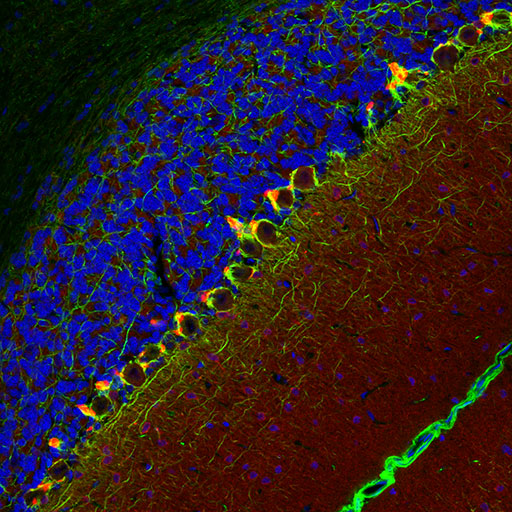
Confocal immunofluorescent analysis of rat cerebellum using PSD95 (D27E11) XP® Rabbit mAb (red) and Neurofilament-L (DA2) Mouse mAb #2835 (green). Blue pseudocolor = DRAQ5 #4084 (fluorescent DNA dye).

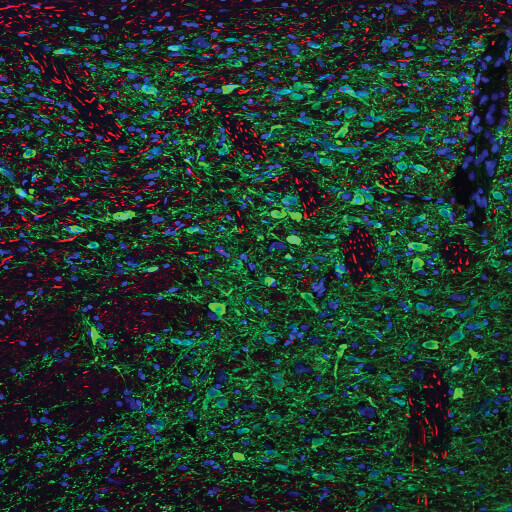
Confocal immunofluorescent analysis of rat substantia nigra shown at low magnification (left) using Tyrosine Hydroxylase (A8Y7R) Rabbit mAb (green) and Neurofilament-L (DA2) Mouse mAb #2835 (red). Blue pseudocolor = DRAQ5 #4084 (fluorescent DNA dye).
The release of neurotransmitters is dependent on changes in intracellular voltage - which is mediated by ligand and gated ion channels in the presynaptic cell. Depolarization of the cell results in action potential propagation through the entire axon. At the presynaptic terminal, calcium influx stimulates the extracellular release of the neurotransmitter vesicles.
After traversing the synapse, neurotransmitters bind to postsynaptic receptors on the dendrites and exert either an excitatory or inhibitory response.
Following the action potential, the presynaptic cell repolarizes using the action of ion channels and ATP-dependent transporters. Neurotransmission is terminated by neurotransmitter enzymatic degradation in the synaptic cleft, transported-mediated recycling to its original axon terminal, or by transporter-mediated astrocytic uptake.